. 2013 Jun;73(6):695-704.
doi: 10.1002/ana.23892. Epub 2013 Jun 5.

Cell age-specific vulnerability of neurons to anesthetic toxicity

Affiliations

Cell age-specific vulnerability of neurons to anesthetic toxicity

Rylon D Hofacer et al. Ann Neurol. 2013 Jun.

Abstract

Objective: Anesthetics have been linked to widespread neuronal cell death in neonatal animals. Epidemiological human studies have associated early childhood anesthesia with long-term neurobehavioral abnormalities, raising substantial concerns that anesthetics may cause similar cell death in young children. However, key aspects of the phenomenon remain unclear, such as why certain neurons die, whereas immediately adjacent neurons are seemingly unaffected, and why the immature brain is exquisitely vulnerable, whereas the mature brain seems resistant. Elucidating these questions is critical for assessing the phenomenon's applicability to humans, defining the susceptible age, predicting vulnerable neuronal populations, and devising mitigating strategies.

Methods: This study examines the effects of anesthetic exposure on late- and adult-generated neurons in newborn, juvenile, and adult mice, and characterizes vulnerable cells using birth-dating and immunohistochemical techniques.

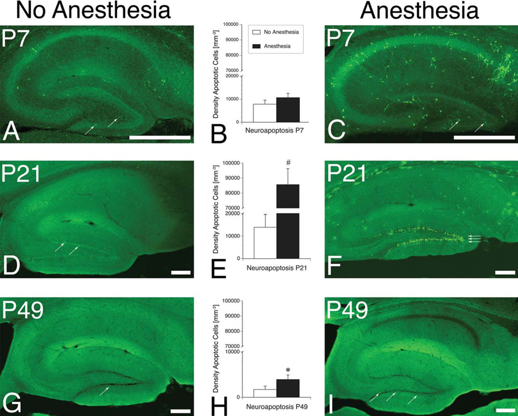
Results: We identify a critical period of cellular developmental during which neurons are susceptible to anesthesia-induced apoptosis. Importantly, we demonstrate that anesthetic neurotoxicity can extend into adulthood in brain regions with ongoing neurogenesis, such as dentate gyrus and olfactory bulb.

Interpretation: Our findings suggest that anesthetic vulnerability reflects the age of the neuron, not the age of the organism, and therefore may potentially not only be relevant to children but also to adults undergoing anesthesia. This observation further predicts differential heightened regional vulnerability to anesthetic neuroapoptosis to closely follow the distinct regional peaks in neurogenesis. This knowledge may help guide neurocognitive testing of specific neurological domains in humans following exposure to anesthesia, dependent on the individual's age during exposure.


Conflict of interest statement

Potential Conflicts of Interest

S.C.D.: travel expenses, Yuying Children Hospital, Wenzhou Medical University.

Figures

FIGURE 1
FIGURE 1
Vulnerability to anesthesia-induced neuroapoptosis is delayed in dentate granule cells and continues into adulthood. Representative hippocampal photomicrographs stained for activated caspase 3 (positive cells are seen as bright green puncta) from newborn (postnatal day 7 [P7]; A, C), juvenile (P21; D, F), and young adult (P49; G, I) mice exposed to 1.5% of isoflurane (anesthesia) for 6 hours, compared with fasted, unanesthetized littermates (no anesthesia). Arrows mark apoptotic dentate granule cells. Bar graphs (B, E, H) represent quantification of apoptotic neuronal density (n = 6–8 animals per group for each time point, mean ± standard error of the mean). Stereological cell counts of caspase 3+ cellular density reveal that neuroapoptosis is not significantly increased, compared with unanesthetized littermates, following anesthetic exposure at P7, but is increased following anesthetic exposure at P21 (note y-axis break) and P49; #p < 0.001, *p < 0.05, Mann–Whitney U test. All scale bars = 200µm. [Color figure can be viewed in the online issue, which is available at wileyonlinelibrary.com]
FIGURE 2
FIGURE 2
Dentate granule cell vulnerability to anesthesia-induced neuroapoptosis is dependent on neuronal age and peaks approximately 2 weeks after the cells are born. Animals were injected with the S-phase marker bromodeoxyuridine (BrdU) at 4 time points: 3 to 5, 8 to 10, 13 to 15, or 18 to 20 days prior to an anesthetic exposure of 1.5% isoflurane for 6 hours on postnatal day 21 (P21) or P49. (A) Graph depicts the fraction of birth-dated cells from each injection point that were vulnerable to anesthesia-induced apoptosis by dividing the number of caspase 3+/BrdU+ double-positive cells by the number of all BrdU+ cells (n = 17–32 animals for each time point; *p = 0.008 compared with cells older than 18 days; other comparisons were not significant, Mann–Whitney U test). (B–D) Representative high-magnification photomicrograph composite images of dentate granule cells from a juvenile mouse following anesthetic exposure shows neurons positive for the apoptotic marker activated caspase 3 (green, B), the S-phase marker BrdU (red, C), and a merged image (D). Arrows mark 2 cells double-positive for caspase 3 and BrdU, as quantified in A. (E–G) Similar representative images from a young adult, P49 mouse, which was injected with BrdU from P39 to P41, identifying the double-labeled neuron (arrows) as being an adult-generated dentate granule cell, between 8 and 10 days old during the anesthetic exposure on P49; note 2 neighboring neurons of similar age (BrdU+), but not colocalizing with caspase 3. Scale bars = 30µm. [Color figure can be viewed in the online issue, which is available at wileyonlinelibrary.com]
FIGURE 3
FIGURE 3
Anesthesia triggers cell death in newly generated dentate granule cells (DGCs), as identified by their location and phenotypic markers, irrespective of the age of the animal. (A–D) Representative photomicrograph composites of dentate gyrus from a 21-day-old (P21) mouse, following an isoflurane anesthetic, stained for the apoptotic marker caspase 3 (green, A), the mature granule cell marker calbindin (red, B), the immature granule cell marker calretinin (blue, C), and a merged image (D). Caspase 3+, apoptotic neurons are predominantly located in the subgranular zone (arrows), and express the immature marker calretinin but not the mature marker calbindin. (E) Bar graphs represent the fraction of apoptotic, caspase 3+ DGCs colocalizing the respective maturational stage-specific markers (caspase 3+ cells colabeled with 1 of the 4 respective markers divided by the total number of caspase 3+ cells). Apoptotic DGCs are identified as late progenitors (349 of 586 caspase+ cells were NeuroD1+; *p < 0.0001 compared with Sox2+ by Pearson chi-square test) and immature granule cells (849 of 1,587 caspase+ cells were calretinin+; #p < 0.0001 compared with calbindin+ by Pearson chi-square test), but not as radial glialike progenitors (0 of 279 caspase+ cells were Sox2+) or mature neurons (11 of 1587 caspase+ cells were calbindin+; n = 18 animals). (F) Schematic demonstrating DGC maturation progressing from radial glialike progenitor cells, located close to the hilus and expressing Sox2, to transient amplifying cells and neuroblasts, located in the subgranular zone (SGZ) and expressing NeuroD1, to immature neurons with small dendritic trees, located in the SGZ and expressing calretinin, and lastly to calbindin-expressing mature neurons migrating into the granule cell layer (GCL) and extending dendrites into the molecular layer (ML). (G–J) Similar composite photomicrographs from a young adult mouse (P49), and (K) bar graphs of characterization identifying apoptotic cells as late progenitors (NeuroD1 [116 of 358]; *p < 0.0001 compared with Sox2+ [0 of 160] by Pearson chi-square test) and immature granule cells (calretinin+ [76 of 288]; #p < 0.0001, compared with calbindin+ [3 of 288] by Pearson chi-square test; n = 16 animals). All scale bars = 50µm. [Color figure can be viewed in the online issue, which is available at wileyonlinelibrary.com]
FIGURE 4
FIGURE 4
Vulnerability to anesthesia-induced neuroapoptosis extends into adulthood in olfactory bulb, another brain region incorporating adult-generated neurons. Representative photomicrographs of olfactory bulbs and quantification of stereological cell counts from unanesthetized, fasted animals, compared with their respective littermates exposed to 1.5% isoflurane for 6 hours on (A–E) postnatal day 7 (P7), (F–J) P21, or (K–O) P49 demonstrating caspase 3+, apoptotic neurons (bright green puncta) and neurons staining for the neuronal marker NeuN (blue areas). White boxes on the left (A, D, F, I, K, N) correspond to respective high-resolution images on the right (B, E, G, J, L, O). Quantification of stereological cell counts of apoptotic neurons in newborn (C), juvenile (H), and young adult mice (M) demonstrate a significant increase of caspase 3+ olfactory bulb neurons (arrows in B, E, G, J, L, O) following anesthetic exposure (#p < 0.01, *p < 0.001, †p < 0.05 compared with their respective, unanesthetized littermates; Mann–Whitney U test; n = 6–8 animals per group). All scale bars = 250µm. [Color figure can be viewed in the online issue, which is available at wileyonlinelibrary.com]

References

    1. Weiser TG, Regenbogen SE, Thompson KD, et al. An estimation of the global volume of surgery: a modelling strategy based on available data. Lancet. 2008;372:139–144. - PubMed
    1. Ikonomidou C, Bosch F, Miksa M, et al. Blockade of NMDA receptors and apoptotic neurodegeneration in the developing brain. Science. 1999;283:70–74. - PubMed
    1. Jevtovic-Todorovic V, Hartman RE, Izumi Y, et al. Early exposure to common anesthetic agents causes widespread neurodegeneration in the developing rat brain and persistent learning deficits. J Neurosci. 2003;23:876–882. - PMC - PubMed
    1. Stefovska VG, Uckermann O, Czuczwar M, et al. Sedative and anticonvulsant drugs suppress postnatal neurogenesis. Ann Neurol. 2008;64:434–445. - PubMed
    1. Brambrink AM, Evers AS, Avidan MS, et al. Isoflurane-induced neuroapoptosis in the neonatal rhesus macaque brain. Anesthesiology. 2010;112:834–841. - PMC - PubMed

Publication types