. 2012 Dec:2012:694137.
doi: 10.6064/2012/694137.

Retroviral and lentiviral vectors for the induction of immunological tolerance

Affiliations

Retroviral and lentiviral vectors for the induction of immunological tolerance

Inès Dufait et al. Scientifica (Cairo). 2012 Dec.

Abstract

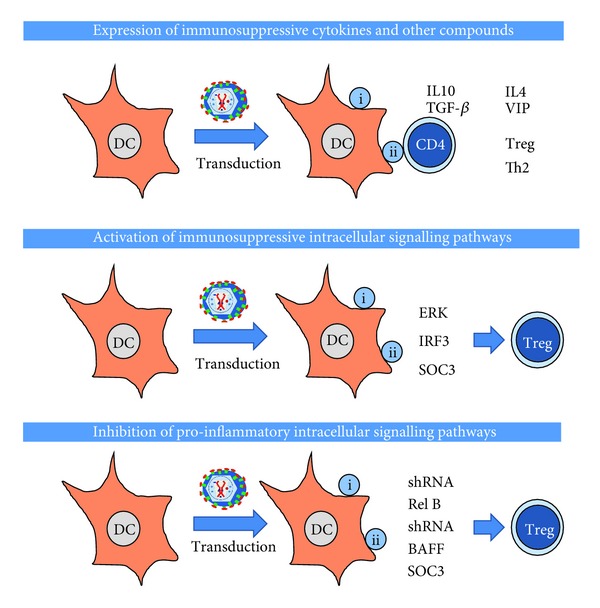
Retroviral and lentiviral vectors have proven to be particularly efficient systems to deliver genes of interest into target cells, either in vivo or in cell cultures. They have been used for some time for gene therapy and the development of gene vaccines. Recently retroviral and lentiviral vectors have been used to generate tolerogenic dendritic cells, key professional antigen presenting cells that regulate immune responses. Thus, three main approaches have been undertaken to induce immunological tolerance; delivery of potent immunosuppressive cytokines and other molecules, modification of intracellular signalling pathways in dendritic cells, and de-targeting transgene expression from dendritic cells using microRNA technology. In this review we briefly describe retroviral and lentiviral vector biology, and their application to induce immunological tolerance.

Keywords: Autoimmune disease; MAPK; NF-κB; T cell; arthritis; co-stimulation; dendritic cell; immunotherapy; microRNA; signalling; tolerance.


Figures

Figure 1
Figure 1
General structure of the retrovirion. The structure of the retrovirion is schematically shown in the figure. The retrovirus contains a diploid RNA genome associated to the nucleocapsid protein (NC), forming the nucleocapsid. This nucleocapsid is associated to structural proteins involved in the retrotranscription (RT), integration (integrase, IN), and virion maturation (protease, PR). All these are enclosed by the capsid (CA) and matrix protein (MA). Then, the virion envelope encloses the core and contains the envelope glycoprotein. In the case of retroviruses and lentiviruses, the envelope protein is made of the transmembrane (TM) and globular subunit (SU).
Figure 2
Figure 2
General life cycle of retrovirus/lentivirus. The lentivirus life cycle is schematically depicted in this Figure. First, the virion can enter the cell either by endocytosis of direct fusion with the cell membrane after binding to its specific receptor (top of the figure). Then, the retrovirus core is released (core release) and reverse transcription takes place. The core containing the cDNA virus genome is transported to the nucleus where it integrates into the host cell chromosome (provirus integration). From the provirus, transcription takes place leading to the transport of either full-length RNA genomes, or to mRNAs encoding the structural and enzymatic proteins (translation). The structural proteins package the full-length RNA genome during virion budding (see right of the figure), releasing infectious viruses following virion maturation.
Figure 3
Figure 3
Engineering of the lentivector gene transfer system. The HIV-1 genome organisation is shown on the top, from 5′ to 3′, long-terminal repeats (LTR), packaging signal (PS), GAG-PRO-POL-In genes, envelope gene (ENV), and the accessory genes Vif, Vpr, Rev, Tat, Nef, and the Rev-response element (RRE). This genome is separated into three different plasmid constructs. The transfer vector (vector), that contains at least, the LTR, PS, and the internal promoter controlling the transcription of the gene of interest. The packaging plasmid, which leads to the expression of the GAG-POL-PRO-IN, rev, and tat genes under the control of the cytomegalovirus (CMV) promoter. Lastly, the envelope plasmid, which expresses the required envelope glycoprotein which will confer the tropism to the vector particle. Cotransfection of these three plasmids will lead to the production of lentivirus-like particles with the capacity of transducing target cells independently of their cell cycle.
Figure 4
Figure 4
Antigen presentation and T cell responses. Activation of T cell responses by antigen presenting dendritic cells is shown in the figure. On the left, a DC is presenting antigenic peptides associated to MHC I (sphere containing “I”) or MHC II (sphere containing “II”) molecules. CD8 or CD4 T cells recognise these peptide-MHC molecules together with additional receptor-ligand interactions (costimulation, represented by bars connecting the DC with each T cell). These interactions accompanied by the presence of a wide range of cytokines will drive T cell proliferation (right). CD8 T cells will then differentiate into cytotoxic T lymphocytes, and CD4 T cells into helper T cells which will collaborate in raising antibody responses. T helper differentiation can lead to either immunological tolerance or different types of immune responses, such as a “Th1-” type (mainly cellular immunity) or a “Th2-” type (mainly humoral immunity).
Figure 5
Figure 5
Main strategies for the induction of immunological tolerance by genetic modification of DCs using retroviral/lentiviral vectors. In this scheme, the most utilised strategies for the induction of immunological tolerance using gene modification of DCs are shown. On top, using lentiviral or retroviral vectors, the expression of immunosuppressive cytokines in DCs can induce the differentiation of antigen-specific Tregs, or in some cases, Th2 cells. In the middle, lentivectors can be used to express constitutive activators or immunosuppressive intracellular signalling pathways leading to the differentiation of suppressive T cells. On the bottom, lentiviral vectors can either deliver short hairpin RNAs targeted towards proinflammatory pathways such as NF-κB, proinflammatory receptors such as BAFF, or inhibitors of cytokine signalling such as SOC3. All these strategies also lead to the development of suppressive T cells. Not shown in any of these schemes, all strategies lead to differentiation of tolerogenic DCs which will either prevent the expansion of cytotoxic CD8 T cells, or induce their apoptosis/anergy.

References

    1. Mann R, Mulligan RC, Baltimore D. Construction of a retrovirus packaging mutant and its use to produce helper-free defective retrovirus. Cell. 1983;33(1):153–159. - PubMed
    1. Anderson WF, Blaese RM, Culver K. The ADA Human Gene Therapy clinical protocol: points to consider response with clinical protocol, July 6, 1990. Human Gene Therapy. 1990;1(3):331–362. - PubMed
    1. Blaese RM, Culver K, Culver K, et al. Treatment of severe combined immunodeficiency disease (SCID) due to adenosine deaminase deficiency with autologous lymphocytes transduced with a human ADA gene. Human Gene Therapy. 1993;4(4):521–527. - PubMed
    1. Cavazzana-Calvo M, Thrasher A, Mavillo F. The future of gene therapy. Nature. 2004;427(6977):779–781. - PubMed
    1. Levine F, Friedmann T. Gene therapy techniques. Current Opinion in Biotechnology. 1991;2(6):840–844. - PubMed

LinkOut - more resources