. 2013;8(3):e50713.
doi: 10.1371/journal.pone.0050713. Epub 2013 Mar 19.

Synthesis and characterization of dual-functionalized core-shell fluorescent microspheres for bioconjugation and cellular delivery

Affiliations

Synthesis and characterization of dual-functionalized core-shell fluorescent microspheres for bioconjugation and cellular delivery

Jonathan M Behrendt et al. PLoS One. 2013.

Abstract

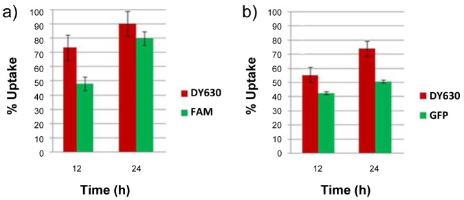
The efficient transport of micron-sized beads into cells, via a non-endocytosis mediated mechanism, has only recently been described. As such there is considerable scope for optimization and exploitation of this procedure to enable imaging and sensing applications to be realized. Herein, we report the design, synthesis and characterization of fluorescent microsphere-based cellular delivery agents that can also carry biological cargoes. These core-shell polymer microspheres possess two distinct chemical environments; the core is hydrophobic and can be labeled with fluorescent dye, to permit visual tracking of the microsphere during and after cellular delivery, whilst the outer shell renders the external surfaces of the microspheres hydrophilic, thus facilitating both bioconjugation and cellular compatibility. Cross-linked core particles were prepared in a dispersion polymerization reaction employing styrene, divinylbenzene and a thiol-functionalized co-monomer. These core particles were then shelled in a seeded emulsion polymerization reaction, employing styrene, divinylbenzene and methacrylic acid, to generate orthogonally functionalized core-shell microspheres which were internally labeled via the core thiol moieties through reaction with a thiol reactive dye (DY630-maleimide). Following internal labeling, bioconjugation of green fluorescent protein (GFP) to their carboxyl-functionalized surfaces was successfully accomplished using standard coupling protocols. The resultant dual-labeled microspheres were visualized by both of the fully resolvable fluorescence emissions of their cores (DY630) and shells (GFP). In vitro cellular uptake of these microspheres by HeLa cells was demonstrated conventionally by fluorescence-based flow cytometry, whilst MTT assays demonstrated that 92% of HeLa cells remained viable after uptake. Due to their size and surface functionalities, these far-red-labeled microspheres are ideal candidates for in vitro, cellular delivery of proteins.


Conflict of interest statement

Competing Interests: The authors have declared that no competing interests exist.

Figures

Figure 1
Figure 1. Synthesis of core-shell microspheres.
Reaction conditions: i) PVP 58k, azobisisobutyronitrile (AIBN), hexadecane, 70°C, 16 h ii) AIBN, hexadecane, SDS(aq), RT 70°C, 5 h.
Figure 2
Figure 2. SEM examination of microspheres.
SEM images of thiouronium-functionalized microspheres 4, core-shell microspheres 6a6c synthesized with varying ratios of co-monomers to seed particles and core shell microspheres 6e and 6f synthesized with varying ratios of styrene to methacrylic acid.
Figure 3
Figure 3. Fluorescent labeling of core-shell microspheres.
Reaction conditions: i) DMF, MeOH, RT, 16 h; ii) DMF, RT, 2 h; iii) EDAC, MES pH 5.5, DMF, RT, 18 h; iv)EDAC, MES pH 6.5, NaOH, RT, 2.5 h.
Figure 4
Figure 4. Flow cytometry analysis of microspheres.
Flow cytometry analysis of a) unlabelled microspheres 7a (negative control); b) DY-630 labelled microspheres 8a; c) DY-630 labelled microspheres 8b
Figure 5
Figure 5. Confocal microscopy of microspheres.
Confocal microscope images of DY-630 labelled microspheres conjugated to GFP 10a: a) excited at 633 nm; b) excited at 488 nm; c) composite image showing co-localization of DY630 and GFP fluorescence. Scale bar = 4 µm.
Figure 6
Figure 6. Confocal microscopy of a single microsphere.
An overlay confocal microscope image of a DY-630 labelled microsphere conjugated to GFP 10a: excited at 633 nm (core, red) and at 488 nm (shell, green). Scale bar = 2 µm.
Figure 7
Figure 7. Cellular uptake of core-labeled microspheres, assessed by FACS.
Percentage cellular uptake of DY-630 labelled microspheres 8 by HeLa cells, as measured by flow cytometry: a) 8a (1 µm diameter); b) 8b (500 nm diameter).
Figure 8
Figure 8. Fluorescent microscopy of beadfected HeLa cells.
Fluorescence microscope images obtained from a Z stack set of images (approximately 0.5 µm z-steps, taken from the top to the bottom focal plane of the cells, 10–12 slices in total) of HeLa cells beadfected with a sample of DY-630-labelled microspheres conjugated via their shells to GFP 10a: a) under irradiation at 633 nm to show DY-630 fluorescence; b) under irradiation at 433 nm to show GFP fluorescence. In each case the cells have been fixed and their nuclei stained with 4',6-diamidino-2-phenylindole (DAPI). Scale bar = 10 µm.
Figure 9
Figure 9. Ceullular uptake of dual core/shell-labeled microspheres, assessed by FACS.
Cellular uptake of shell-conjugated, DY-630 core-labelled microspheres by HeLa cells, as measured by flow cytometry: a) shell conjugated to fluoresceinamine 9a; b) shell conjugated to GFP 10a.

References

    1. Pallen MJ (2011) Time to recognise that mitochondria are bacteria? Trends Microbiol 19: 58–64. - PubMed
    1. Kutschera U, Niklas KJ (2005) Endosymbiosis, cell evolution, and speciation. Theory in Biosciences 124: 1–24. - PubMed
    1. Wang SH, Lee CW, Chiou A, Wei PK (2011) Size-dependent endocytosis of gold nanoparticles studied by three-dimensional mapping of plasmonic scattering images. J Nanobiotechnol 8 Article 33. - PMC - PubMed
    1. Dhar S, Daniel WL, Giljohann DA, Mirkin CA, Lippard SJ (2009) Polyvalent oligonucleotide gold nanoparticle conjugates as delivery vehicles for platinum(IV) warheads. J Am Chem Soc 131: 14652–14653. - PMC - PubMed
    1. Li X, Xie QR, Zhang J, Xia W, Gu H (2011) The packaging of siRNA within the mesoporous structure of silica nanoparticles. Biomaterials 32: 9546–9556. - PubMed

Publication types