. 2013;8(3):e59206.
doi: 10.1371/journal.pone.0059206. Epub 2013 Mar 19.

Molecular modeling of disease causing mutations in domain C1 of cMyBP-C

Affiliations

Molecular modeling of disease causing mutations in domain C1 of cMyBP-C

Poornima Gajendrarao et al. PLoS One. 2013.

Abstract

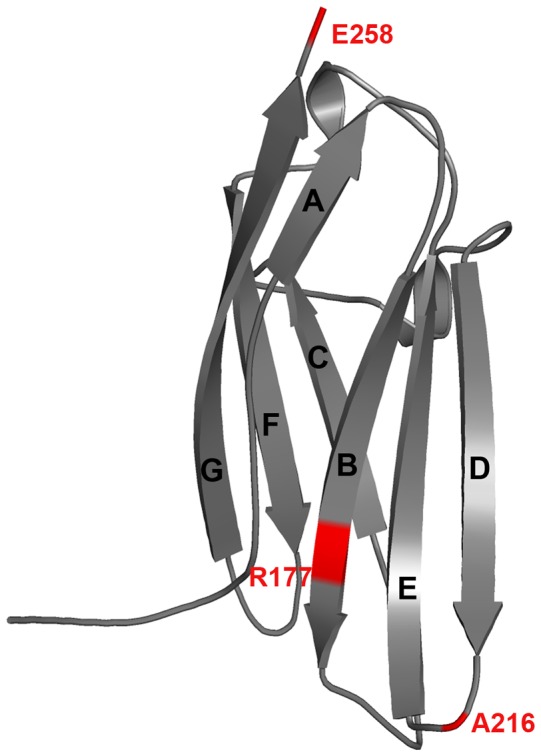
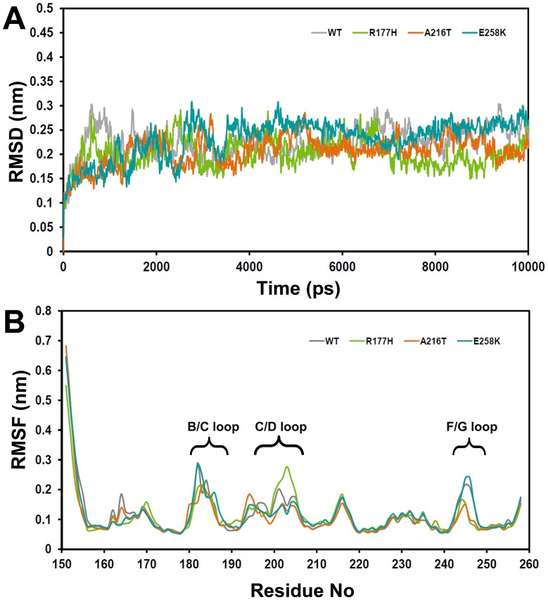
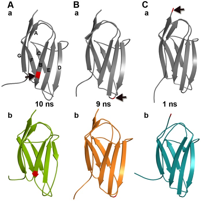
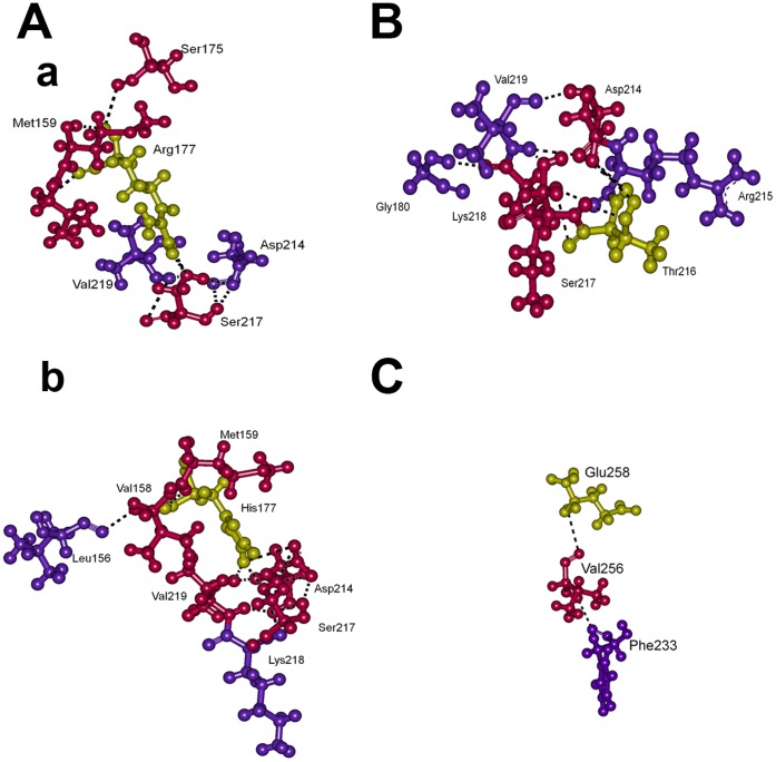
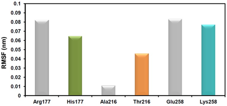
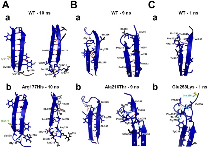
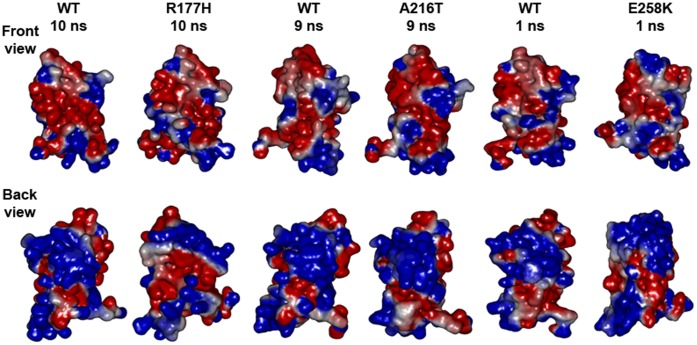
Cardiac myosin binding protein-C (cMyBP-C) is a multi-domain (C0-C10) protein that regulates heart muscle contraction through interaction with myosin, actin and other sarcomeric proteins. Several mutations of this protein cause familial hypertrophic cardiomyopathy (HCM). Domain C1 of cMyBP-C plays a central role in protein interactions with actin and myosin. Here, we studied structure-function relationship of three disease causing mutations, Arg177His, Ala216Thr and Glu258Lys of the domain C1 using computational biology techniques with its available X-ray crystal structure. The results suggest that each mutation could affect structural properties of the domain C1, and hence it's structural integrity through modifying intra-molecular arrangements in a distinct mode. The mutations also change surface charge distributions, which could impact the binding of C1 with other sarcomeric proteins thereby affecting contractile function. These structural consequences of the C1 mutants could be valuable to understand the molecular mechanisms for the disease.


Conflict of interest statement

Competing Interests: The authors have declared that no competing interests exist.

Figures

Figure 1
Figure 1. Structural features of the domain C1 of cMyBP-C.
Positions of the examined HCM causing mutations are shown in red. The C1 consists of seven β-stands that form two β-sheets, where sheet-1 consists of strands A, C, F and G while sheet-2 comprises strands B, D and E both with anti-parallel packing of adjacent strands. Refer Figure 4 and 6 for the key residues.
Figure 2
Figure 2. Trajectory-based structural stability analyses of WT and mutants.
(A) Root mean square deviation (RMSD) and (B) average root mean square fluctuation (RMSF) of residues during MD simulations.
Figure 3
Figure 3. Secondary structural changes.
The representative snapshots of WT and mutants are shown. (A) a) WT and b) Arg177His at 10 ns, (B) a) WT and b) Ala216Thr at 9 ns and (C) a) WT and b) Glu258Lys at 1 ns. In the structures, red color cartoon and black arrows indicate position of the mutation.
Figure 4
Figure 4. Intra-molecular interactions.
For the represented snapshots of MD simulations (Figure 3), two layers of neighbouring residues between corresponding WT and mutant residues along with their hydrogen bonds are displayed. Here, (A) a) WT and b) Arg177His at 10 ns, (B) Ala216Thr at 9 ns and (C) WT at 1 ns. At 9 ns Ala216 of WT and at 1 ns Lys258 of Glu258Lys did not make any interactions, hence they are not shown. WT and mutant residues 177, 216 and 258 are shown in yellow, and the residues directly interacting with these residues are shown in pink and are described as first layer. The residues that are interacting with first layer of residues are depicted as second layer and shown in violet.
Figure 5
Figure 5. Fluctuations of the WT and mutant residues during MD simulations.
The average RMSF of the mutated residues for 10 ns are plotted in 2D bar graph. The bars in gray and in different colours (green, orange and aqua green) represent the WT and the mutants, respectively.
Figure 6
Figure 6. Molecular explanations for the structural changes.
The rigidity analysis provided information based on the structural rigidity and flexibility of residues. (A) 10 ns snapshots of a) WT and b) Arg177His, (B) 9 ns snapshots of a) WT and b) Ala216Thr and (C) 1 ns snapshots of a) WT and b) Glu258Lys. Here, blue represents rigid regions and black and gray indicates flexible regions. The mutational spots are shown in yellow arrow. As the Ala216Thr affected the distal region of the domain, the position of this mutation has not shown. The hydrogen bonds (red lines) that are missing in the mutants are marked in black and the newly formed hydrogen bonds in the mutants are shown in light blue.
Figure 7
Figure 7. Electrostatic surface potential map.
Front view and back view of surface electrostatic potential map for the snapshots shown in Figure 3. Where, blue, red and white represent positive, negative and hydrophobic electrostatic potential, respectively. WT at 10 ns, 9 ns and 1 ns were in different conformational states due to the simulations that were performed after the equilibration. In addition, the N-terminal of the protein was highly flexible during all 4 simulations (Figure 2b), hence the electrostatic surface map of the representative snapshots shows considerable change at their N-terminal.

References

    1. Maron BJ, Gardin JM, Flack JM, Gidding SS, Kurosaki TT, et al. (1995) Prevalence of hypertrophic cardiomyopathy in a general population of young adults. Echocardiographic analysis of 4111 subjects in the CARDIA Study. Coronary Artery Risk Development in (Young) Adults. Circulation 92: 785–789. - PubMed
    1. Maron BJ (2004) Hypertrophic cardiomyopathy: an important global disease. Am J Med 116: 63–65. - PubMed
    1. Maron BJ, Shirani J, Poliac LC, Mathenge R, Roberts WC, et al. (1996) Sudden death in young competitive athletes. Clinical, demographic, and pathological profiles. JAMA 276: 199–204. - PubMed
    1. Cecchi F, Yacoub MH, Olivotto I (2005) Hypertrophic cardiomyopathy in the community: why we should care. Nat Clin Pract Cardiovasc Med 2: 324–325. - PubMed
    1. Marian AJ, Salek L, Lutucuta S (2001) Molecular genetics and pathogenesis of hypertrophic cardiomyopathy. Minerva Med 92: 435–451. - PMC - PubMed

Publication types