

A new non-human primate model of photochemically induced cerebral infarction
- PMID: 23527298
- PMCID: PMC3603910
- DOI: 10.1371/journal.pone.0060037
A new non-human primate model of photochemically induced cerebral infarction
Abstract
Background and purpose: Rat models of photochemically induced cerebral infarction have been readily studied, but to date there are no reports of transcranial photochemically induced infarctions in the marmoset. In this report, we used this non-human primate as a model of cerebral thrombosis and observed the recovery process.
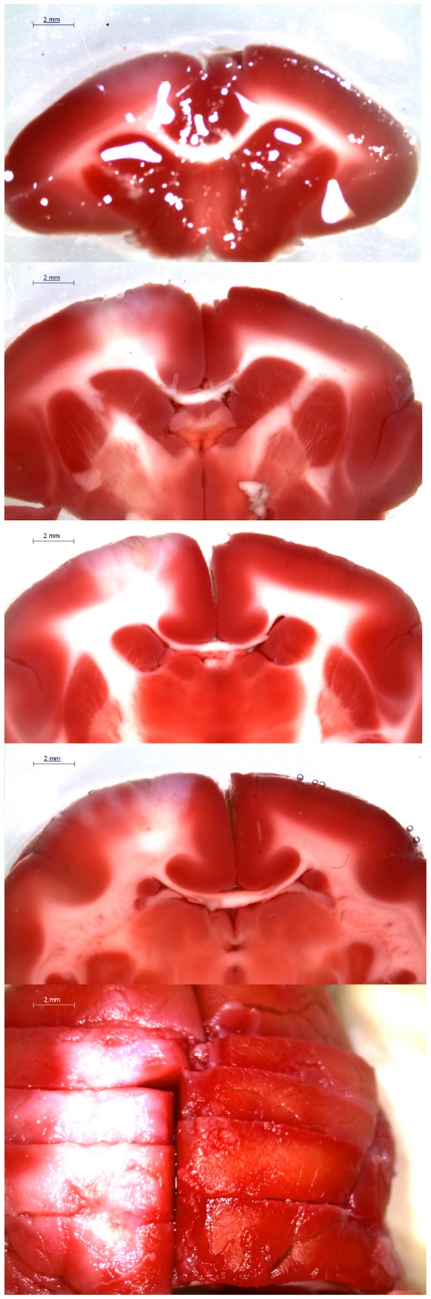
Methods: Five common marmosets were used. Cerebral ischemia was produced via intravascular thrombosis induced by an intravenous injection of Rose Bengal and irradiation with green light. After inducing cerebral infarction, we observed the behavior of marmosets via a continuous video recording. We evaluated maximum speed, mean speed, and distance traveled in 1 min. In addition, we evaluated scores for feeding behavior, upper limb grip, and lower limb grip. We confirmed the infarct area after cerebral infarction using 2,3,5-triphenyltetrazolium chloride staining in a separate marmoset.
Results: We found functional decreases 2 days after creating the cerebral infarction in all measurements. Total distance traveled, average speed, upper limb score, and feeding behavior score did not recover to pre-infarction levels within 28 days. Maximum speed in 1 min and lower limb score recovered 28 days after infarction as compared to pre-infarction levels. We confirmed the infarct area of 11.4 mm × 6.8 mm as stained with 2,3,5-triphenyltetrazolium chloride.
Conclusion: We were able to create a primate photothrombosis-induced cerebral infarction model using marmosets and observe functional recovery. We suggest that this is a useful model for basic research of cerebral infarction.
Conflict of interest statement
Figures


References
-
- Feeney DM, Gonzalez A, Law WA (1982) Amphetamine, haloperidol, and experience interact to affect rate of recovery after motor cortex injury. Science 217: 855–857. - PubMed
-
- Horinouchi K, Ikeda S, Harada K, Ohwatashi A, Kamikawa Y, et al. (2007) Functional recovery and expression of GDNF seen in photochemically induced cerebral infarction. Int J Neurosci 117: 315–326. - PubMed
-
- Bihel E, Roussel S, Toutain J, Bernaudin M, Touzani O (2011) Diffusion tensor MRI reveals chronic alterations in white matter despite the absence of a visible ischemic lesion on conventional MRI: a nonhuman primate study. Stroke 56: 1412–1419. - PubMed
-
- Freret T, Bouet V, Toutain J, Saulnier R, Pro-Sistiaga P, et al. (2008) Intraluminal thread model of focal stroke in the non-human primate. J Cereb Blood Flow Metab 28: 786–796. - PubMed
-
- Marshall JW, Ridley RM (1996) Assessment of functional impairment following permanent middle cerebral artery occlusion in a non-human primate species. Neurodegeneration 5: 275–286. - PubMed
Publication types
MeSH terms
Substances
LinkOut - more resources
Full Text Sources
Other Literature Sources
