Review
. 2013:82:663-92.
doi: 10.1146/annurev-biochem-072909-101058. Epub 2013 Mar 18.

Membrane fission reactions of the mammalian ESCRT pathway

Affiliations
Review

Membrane fission reactions of the mammalian ESCRT pathway

John McCullough et al. Annu Rev Biochem. 2013.

Abstract

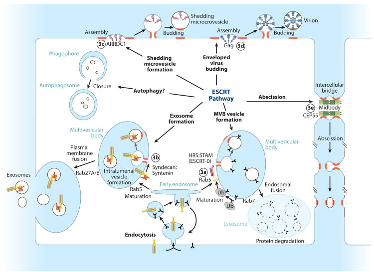
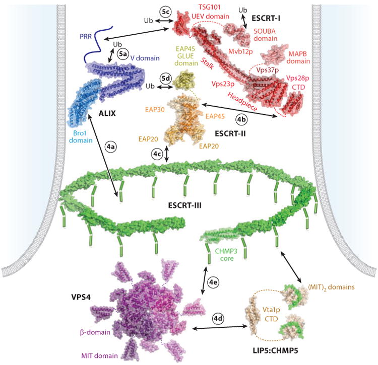
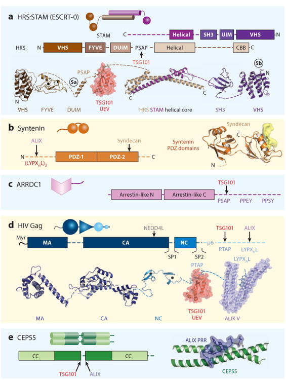
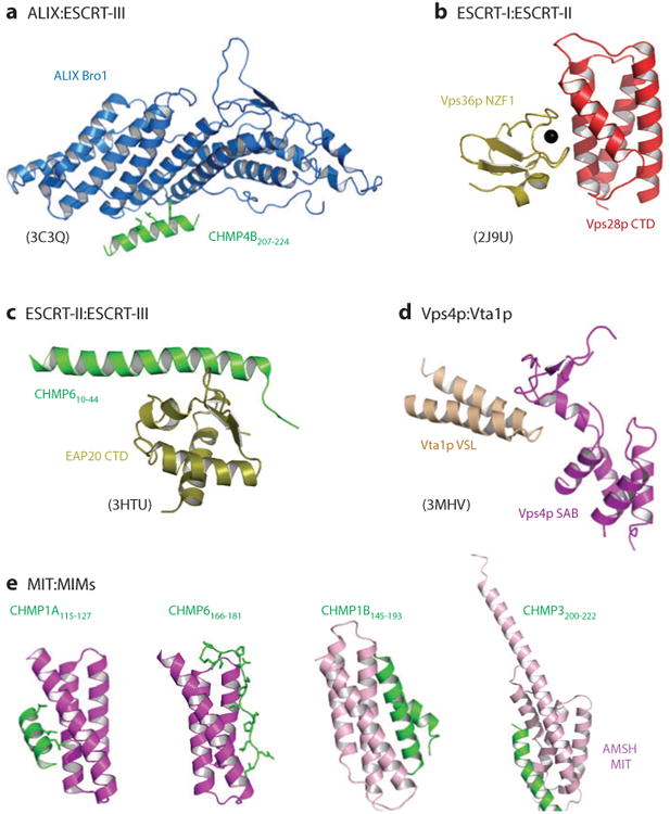
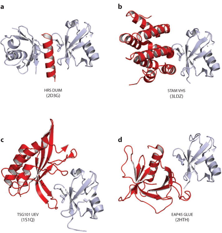
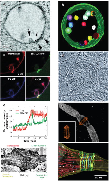
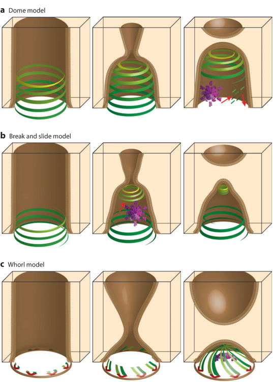
The endosomal sorting complexes required for transport (ESCRT) pathway was initially defined in yeast genetic screens that identified the factors necessary to sort membrane proteins into intraluminal endosomal vesicles. Subsequent studies have revealed that the mammalian ESCRT pathway also functions in a series of other key cellular processes, including formation of extracellular microvesicles, enveloped virus budding, and the abscission stage of cytokinesis. The core ESCRT machinery comprises Bro1 family proteins and ESCRT-I, ESCRT-II, ESCRT-III, and VPS4 complexes. Site-specific adaptors recruit these soluble factors to assemble on different cellular membranes, where they carry out membrane fission reactions. ESCRT-III proteins form filaments that draw membranes together from the cytoplasmic face, and mechanistic models have been advanced to explain how ESCRT-III filaments and the VPS4 ATPase can work together to catalyze membrane fission.


Figures

Figure 1
Figure 1
Membrane fission reactions promoted by the mammalian ESCRT pathway. Adaptor complexes that direct the pathway to specific sites of action are shown schematically, and ESCRT-remodeled membranes are highlighted red. Circled numbers denote the panel in Figure 3 that shows the adaptor structure.
Figure 2
Figure 2
Core ESCRT components and their interactions, shown within a stylized bud neck. Lines denote regions of unknown structure, dashes denote linkers, cylinders denote helices, arrows denote protein-protein interactions, and circled numbers denote the panel in Figure 4 or 5 that shows the relevant interaction. Relevant Protein Data Bank identity numbers are shown in parentheses: human ALIX (20EV); yeast ESCRT-I core (2P22); human TSG101 UEV domain (1S1Q); yeast Vsp28p C-terminal domain (2J9U); MAPB domain (3TOW); SOUBA domain (4AE4), note that the MAPB and SOUBA domains would not be present simultaneously in any single ESCRT-I complex; ESCRT-II core (2ZME); EAP45 GLUE domain (2HTH); human CHMP3 (2GD5), with individual subunits modeled into a circular filament; VPS4 MIT (2JQ9); VPS4 core (1XWI), modeled as a hexamer based on the structure of the related AAA ATPase p97 (1E32); Vta1p C-terminal domain (2RKL); and LIP5:CHMP5 (2LXM). Abbreviations: CTD, C-terminal domain; MIT, microtubule interacting and transport; PRR, proline-rich region; Ub, ubiquitin.
Figure 3
Figure 3
ESCRT adaptors. The functions and cellular locations of the different adaptors are shown in Figure 1. Here, each adaptor is shown as a cartoon representation, as a schematic model showing domain and motif maps, and, where possible, a composite of structurally characterized domains (ribbon diagrams) and intermolecular complexes (surface renderings). Circled numbers in panel a denote ubiquitin complexes that are shown in Figure 5. Relevant Protein Data Bank identity numbers are shown in parentheses. Panel a contains the HRS:STAM helical core (3F1I), HRS tandem VHS-FYVE domain (1DVP), HRS DUIM (2D3G), HRS PSAP:TSG101 UEV domain complex (3OBQ), STAM SH3 domain (1UJ0), and the STAM VHS domain (3LDZ). Panel b contains the syndecan peptide:syntenin PDZ2 domain complex (1YBO). Panel c contains HIV Gag Myr-MA (1UPH), CA (3H47), NC (1MFS), PTAP:TSG101 UEV domain complex (1M4Q), LYPXnL:ALIX (2R05). Panel d contains the CEP55 coiled coil:ALIX proline-rich region (PRR) complex (3E1R). Abbreviation: CBB, clathrin-binding box.
Figure 4
Figure 4
The structural basis for intercomplex interactions within the ESCRT pathway. The relevant interactions are as depicted in Figure 2, except that interacting domains from yeast Vps28p (ESCRT-I) and Vps36p (ESCRT-II) are shown in panel b because the divergent human ESCRT-I:ESCRT-II interaction site has not been structurally characterized and that interacting domains from yeast Vta1p and Vps4p are shown in panel d because the homologous human LIP5:VPS4 interaction has not been structurally characterized. Multiple ESCRT-III MIM:MIT domain interactions have been structurally characterized, and four distinct classes of interactions are shown [and see Figure 2 for the structure of the LIP5(MIT)2:CHMP5 complex]. Relevant Protein Data Bank identity numbers are shown in parentheses. Abbreviations: MIT, microtubule interacting and transport; MIM, MIT interaction motif.
Figure 5
Figure 5
Structurally characterized ubiquitin interactions within the ESCRT pathway. Ubiquitin (Ub)-binding domains from the HRS:STAM adaptor (ESCRT-0) (see Figure 3a) and from ESCRT-I and ESCRT-II (Figure 2) are shown in red, bound to ubiquitin in gray. The Ub I44 side chains are shown explicitly (stick) to emphasize that the very different ubiquitin-binding domains recognize the same Ub surface. Relevant Protein Data Bank identity numbers are provided within the figure.
Figure 6
Figure 6
(a-c) ESCRT pathway functions in multivesicular body (MVB) vesicle formation, (d-e) enveloped virus budding, and (f-h) the abscission stage of cytokinesis. (a) Slice from an electron microscopy (EM) tomogram showing a vesicle budding into an endosome (large arrow) at a site adjacent to the HRS:STAM:clathrin coat (arrowheads). The small arrow shows a gold particle used to label the endosome. (b) EM tomographic reconstruction of an MVB, pseudocolored to show the internal vesicles and limiting membrane (green). Panels a and b were reprinted with permission from Reference 193, copyright 2003, National Academy of Sciences, U.S.A. (c) Fluorescence micrographs showing that Snf7p (CHMP4, green) concentrates at the neck of an MVB-like vesicle budding into a giant unilamellar vesicle in a reconstituted system. Ubiquitin cargos (blue) and membranes (red) are shown to define the vesicle. Reprinted with permission from Reference 15. (d) Cryoelectron microscopy (cryo-EM) image of a budding HIV-1 viral particle. The virion is ∼100 nm in diameter, the Gag protein lattice is visible as a protein-dense layer inside the plasma membrane, and the open neck of the virion is the site where ESCRT-mediated membrane fission occurs. Reprinted with permission from Reference 109, copyright 2012, Cold Spring Harbor Laboratory Press. (e) Graph showing the time course of CHMP4B (ESCRT-III) recruitment (green) as HIV Gag molecules (red) assemble into a single budding virion on a HeLa cell plasma membrane. Note that Gag assembles gradually (over ∼7 min), whereas CHMP4B appears in a sharp burst immediately prior to virion release. Reprinted with permission from Reference 92. (f) Structured illumination microscopy fluorescence image of an intercellular bridge prior to abscission, showing TSG101 (orange) forming two rings on either side of the midbody (with an alternate view inset) and microtubules (white). Reprinted with permission from Reference 94 (g) Cryo-EM tomographic image of an intercellular bridge showing the midbody, microtubules, and 17-nm filaments within constriction zones that undergo microtubule severing and abscission. Panels g and h were modified and reprinted with permission from Reference 171. (h) Pseudocolored EM reconstructions showing an intercellular bridge late in cytokinesis. Microtubules are shown in red with balls denoting their ends, and 17-nm filaments are shown in green shades. Abscission occurs at the narrow, microtubule-free constriction zone to the right of the filaments.
Figure 7
Figure 7
Models for ESCRT-mediated membrane fission reactions. (a) The “dome” model for multivesicular body (MVB) vesicle budding (188), (b) the “break and slide” model for intercellular bridge constriction and abscission (94, 170), and (c) the “whorl” model for MVB vesicle budding (63). ESCRT-I:ESCRT-II supercomplexes are shown in red; ESCRT-III filaments are shown in shades of green and yellow; VPS4 enzyme complexes are shown in purple; and red arrows denote motion. Panels in the left column show ESCRT factors assembling within membrane tubules (brown). Panels in the central column show models for membrane constriction. Panels in the right column show models for the membrane fission step (see text for details).

References

    1. Henne WM, Buchkovich NJ, Emr SD. The ESCRT pathway. Dev Cell. 2011;21:77–91. - PubMed
    1. Bryant NJ, Stevens TH. Vacuole biogenesis in Saccharomyces cerevisiae: protein transport pathways to the yeast vacuole. Microbiol Mol Biol Rev. 1998;62:230–47. - PMC - PubMed
    1. Raiborg C, Stenmark H. Hrs and endocytic sorting of ubiquitinated membrane proteins. Cell Struct Funct. 2002;27:403–8. - PubMed
    1. Clague MJ, Urbé S. Hrs function: Viruses provide the clue. Trends Cell Biol. 2003;13:603–6. - PubMed
    1. Baietti MF, Zhang Z, Mortier E, Melchior A, Degeest G, et al. Syndecan-syntenin-ALIX regulates the biogenesis of exosomes. Nat Cell Biol. 2012;14:677–85. - PubMed

Publication types

MeSH terms

Substances