. 2013 Mar 19;104(6):1304-13.
doi: 10.1016/j.bpj.2013.01.048. Epub 2013 Mar 19.

Self-assembly and conformational heterogeneity of the AXH domain of ataxin-1: an unusual example of a chameleon fold

Affiliations

Self-assembly and conformational heterogeneity of the AXH domain of ataxin-1: an unusual example of a chameleon fold

Cesira de Chiara et al. Biophys J. .

Abstract

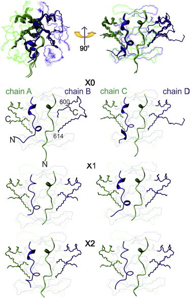
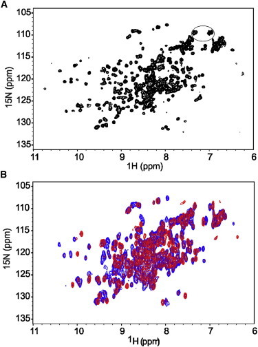
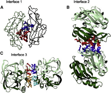
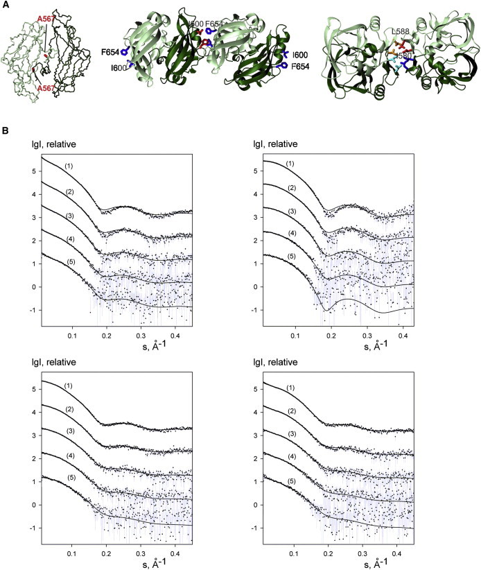
Ataxin-1 is a human protein responsible for spinocerebellar ataxia type 1, a hereditary disease associated with protein aggregation and misfolding. Essential for ataxin-1 aggregation is the anomalous expansion of a polyglutamine tract near the protein N-terminus, but the sequence-wise distant AXH domain modulates and contributes to the process. The AXH domain is also involved in the nonpathologic functions of the protein, including a variety of intermolecular interactions with other cellular partners. The domain forms a globular dimer in solution and displays a dimer of dimers arrangement in the crystal asymmetric unit. Here, we have characterized the domain further by studying its behavior in the crystal and in solution. We solved two new structures of the domain crystallized under different conditions that confirm an inherent plasticity of the AXH fold. In solution, the domain is present as a complex equilibrium mixture of monomeric, dimeric, and higher molecular weight species. This behavior, together with the tendency of the AXH fold to be trapped in local conformations, and the multiplicity of protomer interfaces, makes the AXH domain an unusual example of a chameleon protein whose properties bear potential relevance for the aggregation properties of ataxin-1 and thus for disease.


Figures

Figure 1
Figure 1
Comparison between the crystal structures of the AXH domain of Ataxin-1. (Top) Overlay of the six dimers from the three structures solved under different crystallization conditions and the same bundle rotated by 90°. (Green and blue) Alternated protomers. (Bottom) Individual comparison of the dimers with the chameleon sequence (shown as stick representations).
Figure 2
Figure 2
The NMR spectrum of wt-AXH indicates sample heterogeneity. (A) 15N-1H HSQC spectrum of wt-AXH showing spectral complexity recorded on a 0.40 mM protein solution at 27°C on an Avance spectrometer (Bruker) operating at 700 MHz. (B) Concentration dependence of 15N-1H HSQC spectra of wt-AXH at 0.40 mM (blue) and 0.012 mM (red). Note that the spectra in panel B were collected at lower resolution than the one in panel A (64 vs. 128 increments) to reduce the recording time of the diluted sample. This explains their overall broader appearance. The resonances of glutamines/asparagines are circled.
Figure 3
Figure 3
Cartoon representations of the structural interfaces. (A) Monomer-monomer interface 1 which comprises the first ∼19 residues of protomers A and B of structure X1. (B) Interface 2 between dimers using the same orientation shown in Fig. 2. (Red and blue) Residues from protomers B and C, respectively. (C) Interface 3 between dimers. (Different shades of red and blue) Side chains from different protomers.
Figure 4
Figure 4
SAXS data for wt-AXH and its mutants. (A) Mapping the mutations on the structure. The side chains of the mutated residues are indicated. The orientations are the same used in Fig. 3, A, B, and C. (B) (Top panel) wt-AXH (left) and wt-AXH freshly prepared (right). (Bottom panel) A567G-AXH (left) and I580A-AXH (right). (Curves 1–5) Solute concentrations 1.35, 0.67, 0.33, 0.17, and 0.08 mM, respectively, in 20 mM Tris-HCl pH 7, 2 mM TCEP. (Dots with error bars) Experimental data. (Solid lines) Best fits calculated by the software OLIGOMER (Konarev et al. (28)) for mixtures of monomers, dimers, and tetramers (in the case of wt-AXH and I580A-AXH, higher-order oligomers, i.e., extended octamers and hexadecamers, were taken into account). The intensities are displayed as a function of the momentum transfer and successive curves are displaced down by one logarithmic unit for clarity.
Figure 5
Figure 5
The effect of mutations on the spectral complexity. 15N-1H HSQC spectra of A567G-AXH (left) and of I580A-AXH (right). The spectra were acquired at 0.4 mM protein concentrations and 27°C on an Avance spectrometer (Bruker) operating at 700 MHz. The resonances of asparagines/glutamines are circled.
Figure 6
Figure 6
Further analysis of the spectral complexity. (A) Representative strips from a HNCA spectrum showing at least duplication of the glycine resonances. (B) T1 and T2 values for all peaked resonances. The spectra were recorded on a 0.4 mM protein solution at 27°C on a Varian-INOVA spectrometer operating at 600 MHz.

Similar articles

Cited by

References

    1. Orr H.T., Chung M.Y., Zoghbi H.Y. Expansion of an unstable trinucleotide CAG repeat in spinocerebellar ataxia type 1. Nat. Genet. 1993;4:221–226. - PubMed
    1. Zoghbi H.Y., Orr H.T. Pathogenic mechanisms of a polyglutamine-mediated neurodegenerative disease, spinocerebellar ataxia type 1. J. Biol. Chem. 2008;284:7425–7429. - PMC - PubMed
    1. Matilla-Dueñas A., Goold R., Giunti P. Clinical, genetic, molecular, and pathophysiological insights into spinocerebellar ataxia type 1. Cerebellum. 2008;7:106–114. - PubMed
    1. Chiti F., Dobson C.M. Protein misfolding, functional amyloid, and human disease. Annu. Rev. Biochem. 2006;75:333–366. - PubMed
    1. Soto C., Estrada L.D. Protein misfolding and neurodegeneration. Arch. Neurol. 2008;65:184–189. - PubMed

Publication types

LinkOut - more resources