Comparative Study
. 2013 May 10;288(19):13481-92.
doi: 10.1074/jbc.M112.444810. Epub 2013 Mar 24.

Comparative proteomic analysis of normal and collagen IX null mouse cartilage reveals altered extracellular matrix composition and novel components of the collagen IX interactome

Affiliations
Comparative Study

Comparative proteomic analysis of normal and collagen IX null mouse cartilage reveals altered extracellular matrix composition and novel components of the collagen IX interactome

Bent Brachvogel et al. J Biol Chem. .

Abstract

Background: Collagen IX is an integral cartilage extracellular matrix component important in skeletal development and joint function.

Results: Proteomic analysis and validation studies revealed novel alterations in collagen IX null cartilage.

Conclusion: Matrilin-4, collagen XII, thrombospondin-4, fibronectin, βig-h3, and epiphycan are components of the in vivo collagen IX interactome.

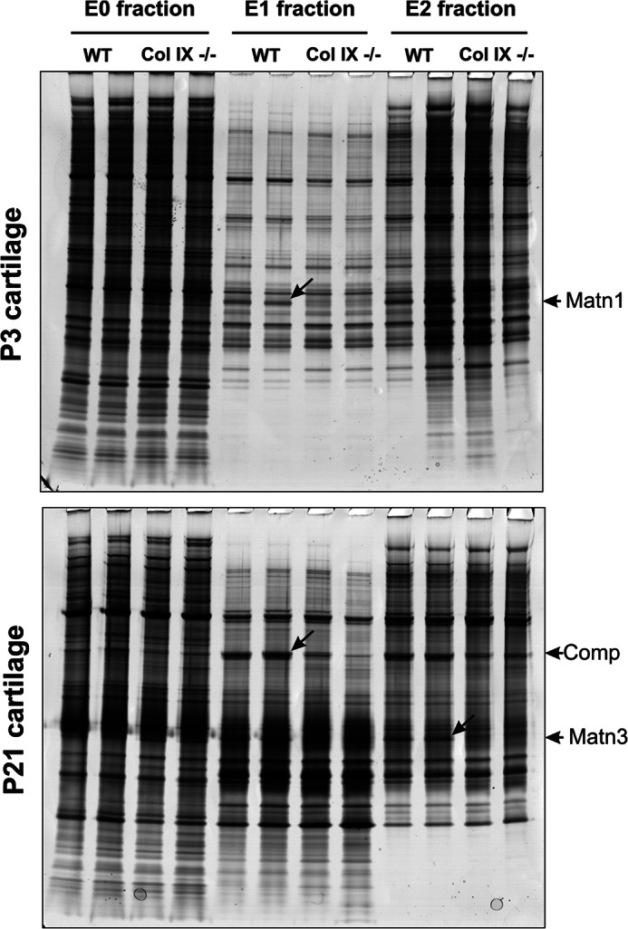
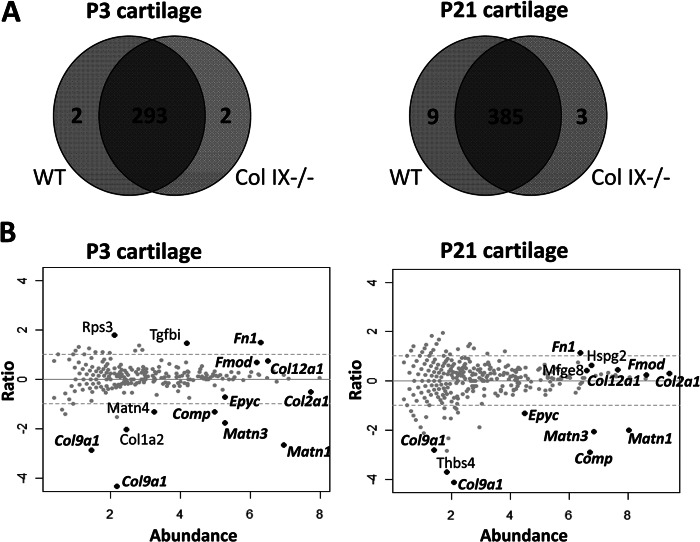
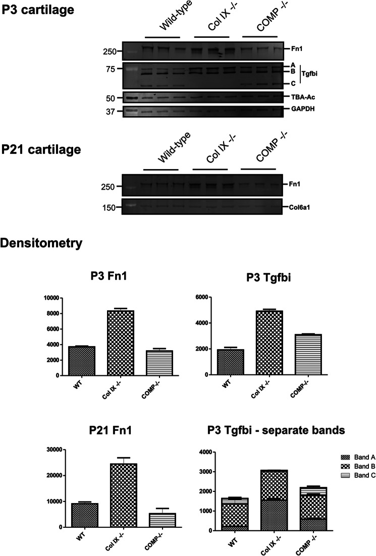
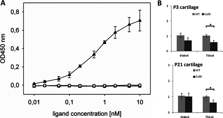
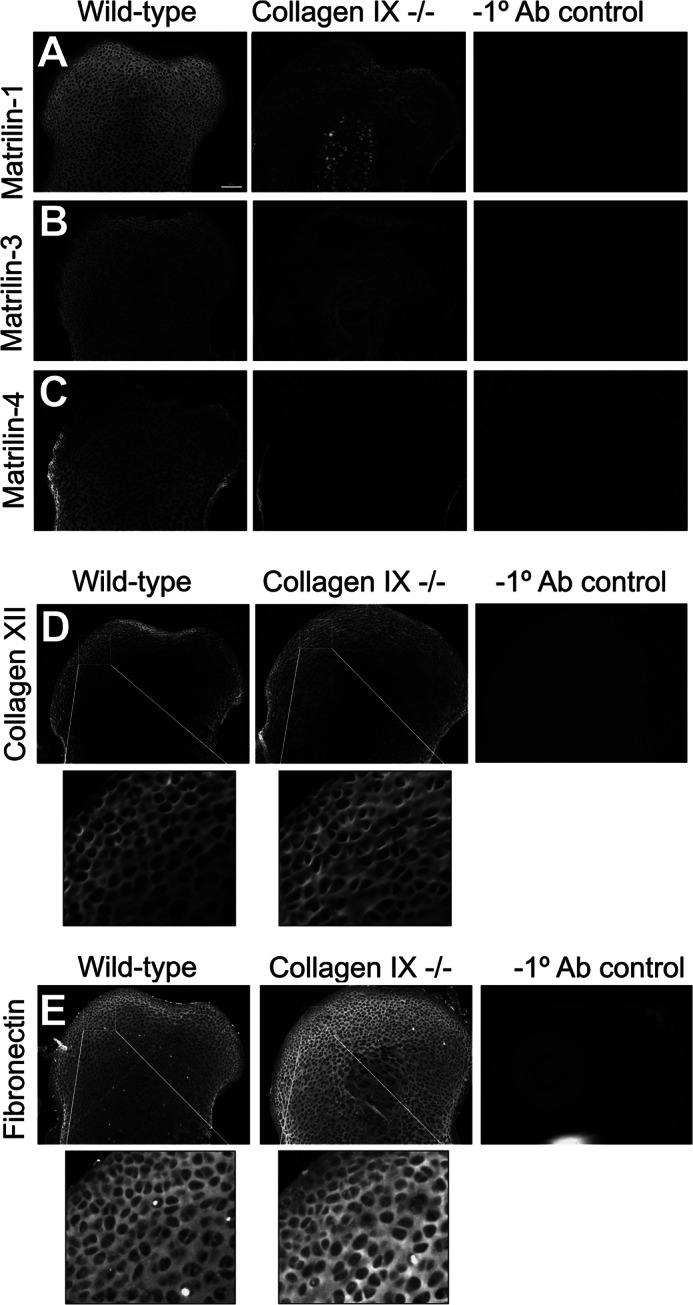
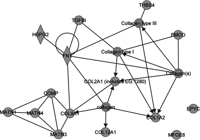
Significance: We applied a proteomics approach to advance our understanding of collagen IX ablation in cartilage. The cartilage extracellular matrix is essential for endochondral bone development and joint function. In addition to the major aggrecan/collagen II framework, the interacting complex of collagen IX, matrilin-3, and cartilage oligomeric matrix protein (COMP) is essential for cartilage matrix stability, as mutations in Col9a1, Col9a2, Col9a3, Comp, and Matn3 genes cause multiple epiphyseal dysplasia, in which patients develop early onset osteoarthritis. In mice, collagen IX ablation results in severely disturbed growth plate organization, hypocellular regions, and abnormal chondrocyte shape. This abnormal differentiation is likely to involve altered cell-matrix interactions but the mechanism is not known. To investigate the molecular basis of the collagen IX null phenotype we analyzed global differences in protein abundance between wild-type and knock-out femoral head cartilage by capillary HPLC tandem mass spectrometry. We identified 297 proteins in 3-day cartilage and 397 proteins in 21-day cartilage. Components that were differentially abundant between wild-type and collagen IX-deficient cartilage included 15 extracellular matrix proteins. Collagen IX ablation was associated with dramatically reduced COMP and matrilin-3, consistent with known interactions. Matrilin-1, matrilin-4, epiphycan, and thrombospondin-4 levels were reduced in collagen IX null cartilage, providing the first in vivo evidence for these proteins belonging to the collagen IX interactome. Thrombospondin-4 expression was reduced at the mRNA level, whereas matrilin-4 was verified as a novel collagen IX-binding protein. Furthermore, changes in TGFβ-induced protein βig-h3 and fibronectin abundance were found in the collagen IX knock-out but not associated with COMP ablation, indicating specific involvement in the abnormal collagen IX null cartilage. In addition, the more widespread expression of collagen XII in the collagen IX-deficient cartilage suggests an attempted compensatory response to the absence of collagen IX. Our differential proteomic analysis of cartilage is a novel approach to identify candidate matrix protein interactions in vivo, underpinning further analysis of mutant cartilage lacking other matrix components or harboring disease-causing mutations.

Keywords: Cartilage; Chondrocytes; Chondrodysplasia; Collagen; Extracellular Matrix; Mass Spectrometry (MS); Proteomics.


Figures

FIGURE 1.
FIGURE 1.
Analysis of sequential extracts of P3 and P21 mouse femoral head cartilage by SDS-PAGE and identification of differentially abundant proteins. Cartilage proteins were partitioned into three fractions based on differential solubility and molecular mass. The 1 m NaCl extract (E0 fraction) was not partitioned further, whereas the 4 m GdnHCl extract was separated into nominal high and low mass E1 and E2 fractions. Aliquots of replicate sequential extracts equivalent to 2% of the protein yield were resolved by 4–12% NuPAGE. Proteins that were clearly different between wild-type (WT) and collagen IX null (Col IX−/−) cartilage extracts, marked by diagonal arrows, were excised, digested with trypsin, and identified by tandem mass spectrometry on an HPLC-interfaced Agilent XCT Plus 3-dimensional ion trap. The differentially abundant proteins were identified as matrilin-1 (Matn1), cartilage oligomeric matrix protein (Comp), and matrilin-3 (Matn3) as indicated by arrows at the gel right-hand margin.
FIGURE 2.
FIGURE 2.
LTQ-Orbitrap analysis of proteins extracted from wild-type and collagen IX-deficient cartilage. A, representation of the proteins identified in P3 and P21 cartilage. The Venn diagrams illustrate the overlap in proteins identified in wild-type (WT) and collagen IX null (Col IX−/−) cartilage extracts from P3 and P21 femoral heads. The numbers of proteins identified with two or more peptide sequences are indicated. B, relative abundance of the proteins identified in wild-type (WT) and collagen IX-deficient (NULL) cartilage. Spectral count data for each protein is plotted using the x axis to represent abundance (½ × log2 [NULL × WT]) and the y axis for fold-change (log2 [NULL/WT]). Proteins increased or decreased in abundance in collagen IX-deficient cartilage are represented by data points above or below the y axis, respectively. Black data points represent the proteins that are significantly altered between the genotypes and are annotated using official gene symbols. Proteins that are differentially abundant in both P3 and P21 cartilage are labeled in bold italics.
FIGURE 3.
FIGURE 3.
Differential two-dimensional electrophoresis analysis of guanidine-extracted proteins from wild-type, collagen IX-deficient, and COMP-deficient cartilage. The ECM-enriched (E1) fraction of wild-type (WT), collagen IX-deficient (Col IX−/−), and COMP-deficient (COMP−/−) cartilage was resolved by two-dimensional electrophoresis, silver stained, and the pseudo-colored images aligned and overlaid using Delta 2D software (Decodon). Proteins that showed clear and consistent genotype-specific differences were identified by tandem mass spectrometry. These proteins, epiphycan (Epyc), matrilin-1 (Matn1), matrilin-3 (Matn3), and hemoglobin (Hbb1), were labeled with arrows. Matn3 is annotated in parentheses to indicate this protein was identified on the basis of a single peptide. The annotation of the COMP protein spot is based on matched migration with COMP previously identified by two-dimensional electrophoresis and mass spectrometry (17). A, proteins more abundant in wild-type cartilage appear as red protein spots and proteins more abundant in collagen IX-deficient cartilage appear as green spots. B, proteins more abundant in wild-type cartilage appear as green protein spots and proteins more abundant in COMP-deficient cartilage appear as red spots.
FIGURE 4.
FIGURE 4.
Validation of proteomics data by fluorescent Western blotting and densitometry. GdnHCl-soluble extracts (E2 fraction) of P3 and P21 wild-type, Col IX−/−, and COMP−/− mouse femoral head cartilage were resolved by SDS-PAGE and probed with antibodies to the TGFβ-induced protein Tgfbi, fibronectin (Fn1). In P21 samples collagen VI (Col6a1) was used as a loading control, as similar levels were observed in the two-dimensional electrophoresis comparison of wild-type, collagen IX-deficient, and COMP-deficient P21 cartilage. In P3 cartilage, GAPDH, and acetylated α-tubulin (TBA-Ac) were used as housekeeping proteins. Background-subtracted volumes for each protein were calculated by densitometry, normalized to the values obtained for the housekeeping proteins to correct for minor loading differences between biological replicates, and plotted as mean values (n = 3) on the y axis, where error bars are mean ± S.E.
FIGURE 5.
FIGURE 5.
Investigation of collagen IX protein binding properties and the relative expression of thrombospondin-4 and matrilin-4 mRNA in wild-type and collagen IX-deficient cartilage. A, binding of soluble matrilin-4 (closed triangles) and thrombospondin-4 (closed squares), to immobilized collagen IX (500 ng/well) was determined using ELISA-style solid phase assays. Uncoated but blocked wells were used as negative controls (open triangles). The two ligands were used at the concentrations indicated and matrilin-4 or thrombospondin-4 specific antibodies were used to detect the level of bound ligand. Duplicate measurements were performed at each concentration and the mean values of seven independent assays for matrilin-4 and three independent assays for thrombospondin-4 are plotted. The resulting saturation curve for matrilin-4 was used to calculate an approximate Kd value of 0.5 nm. B, relative expression level of matrilin-4 (matn4) and thrombospondin-4 (thbs4) in femoral head cartilage of P3 and P21 wild-type and collagen IX-deficient cartilage was determined by qPCR. Results are shown relative to wild-type mRNA expression levels using β actin for normalization. Measurements were performed in triplicate on each biological replicate (n = 6) and the error bars represent the mean ± S.E. Significant differences between the expression levels detected in wild-type and collagen IX were determined using the unpaired two-tailed Student's t test (* = p < 0.05).
FIGURE 6.
FIGURE 6.
Region-specific changes in matrix protein distribution analyzed by immunofluorescence. The protein distribution of matrilin-1 (A), matrilin-3 (B), matrilin-4 (C), collagen XII (D), and fibronectin (E) in the femoral head cartilage of wild-type and collagen IX-deficient P3 mice was determined by immunostaining using in-plane matched paraffin sections. Images from each antibody series, including wild-type negative controls lacking primary antibody were captured using identical microscope and software settings. The boxed regions in D and E were magnified digitally to highlight region-specific differences in staining intensity. Images are representative of biological replicates of wild-type and collagen IX-deficient mice (n = 5). Scale bars, 100 μm.
FIGURE 7.
FIGURE 7.
Interaction network generated using the 15 ECM proteins identified as differentially abundant between collagen IX knock-out and wild-type cartilage. Ingenuity pathway analysis was used to identify relationships between the 15 differentially abundant ECM proteins. In the network, proteins are represented using nodes and relationships between two proteins are represented using edges, where an edge represents a protein-protein interaction, and edges with arrowheads represent membership to a group or complex. All edges are supported by at least one literature reference. The gene products belonging to the collagen protein family are represented by an IPA network in supplemental Fig. S2.

References

    1. Newman B., Wallis G. A. (2003) Skeletal dysplasias caused by a disruption of skeletal patterning and endochondral ossification. Clin. Genet. 63, 241–251 - PubMed
    1. Warman M. L., Cormier-Daire V., Hall C., Krakow D., Lachman R., LeMerrer M., Mortier G., Mundlos S., Nishimura G., Rimoin D. L., Robertson S., Savarirayan R., Sillence D., Spranger J., Unger S., Zabel B., Superti-Furga A. (2011) Nosology and classification of genetic skeletal disorders. 2010 revision. Am. J. Med. Genet. A. 155A, 943–968 - PMC - PubMed
    1. Eyre D. R., Weis M. A., Wu J. J. (2006) Articular cartilage collagen. An irreplaceable framework? Eur. Cell. Mater. 12, 57–63 - PubMed
    1. Briggs M. D., Chapman K. L. (2002) Pseudoachondroplasia and multiple epiphyseal dysplasia. Mutation review, molecular interactions, and genotype to phenotype correlations. Hum. Mutat. 19, 465–478 - PubMed
    1. Bönnemann C. G., Cox G. F., Shapiro F., Wu J. J., Feener C. A., Thompson T. G., Anthony D. C., Eyre D. R., Darras B. T., Kunkel L. M. (2000) A mutation in the α3 chain of type IX collagen causes autosomal dominant multiple epiphyseal dysplasia with mild myopathy. Proc. Natl. Acad. Sci. U.S.A. 97, 1212–1217 - PMC - PubMed

Publication types

MeSH terms

LinkOut - more resources