. 2013 Jun;33(11):2202-11.
doi: 10.1128/MCB.01004-12. Epub 2013 Mar 25.

Farnesoid X receptor inhibits the transcriptional activity of carbohydrate response element binding protein in human hepatocytes

Affiliations

Farnesoid X receptor inhibits the transcriptional activity of carbohydrate response element binding protein in human hepatocytes

Sandrine Caron et al. Mol Cell Biol. 2013 Jun.

Abstract

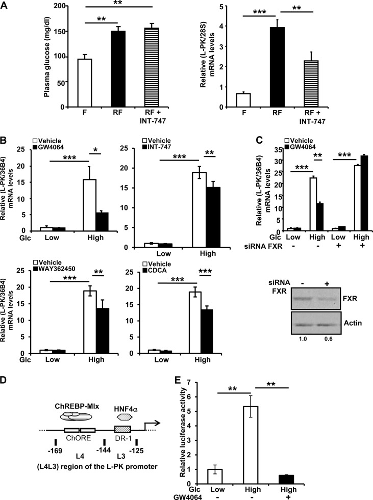
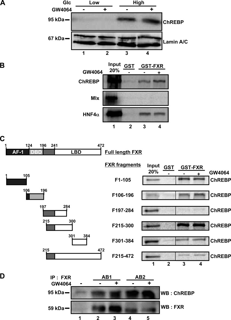
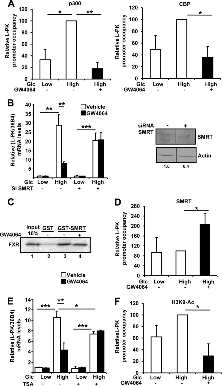
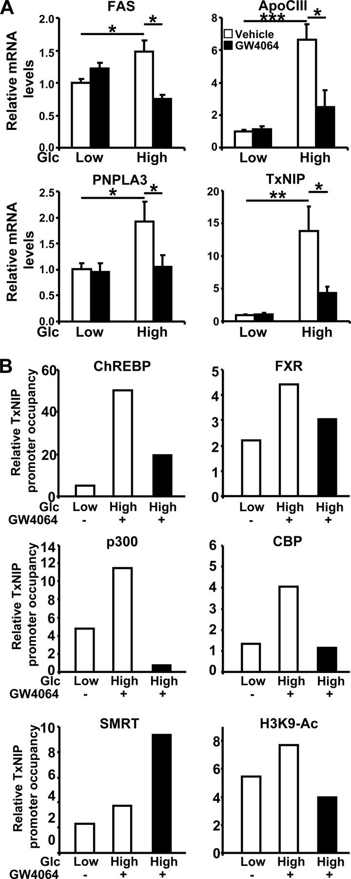
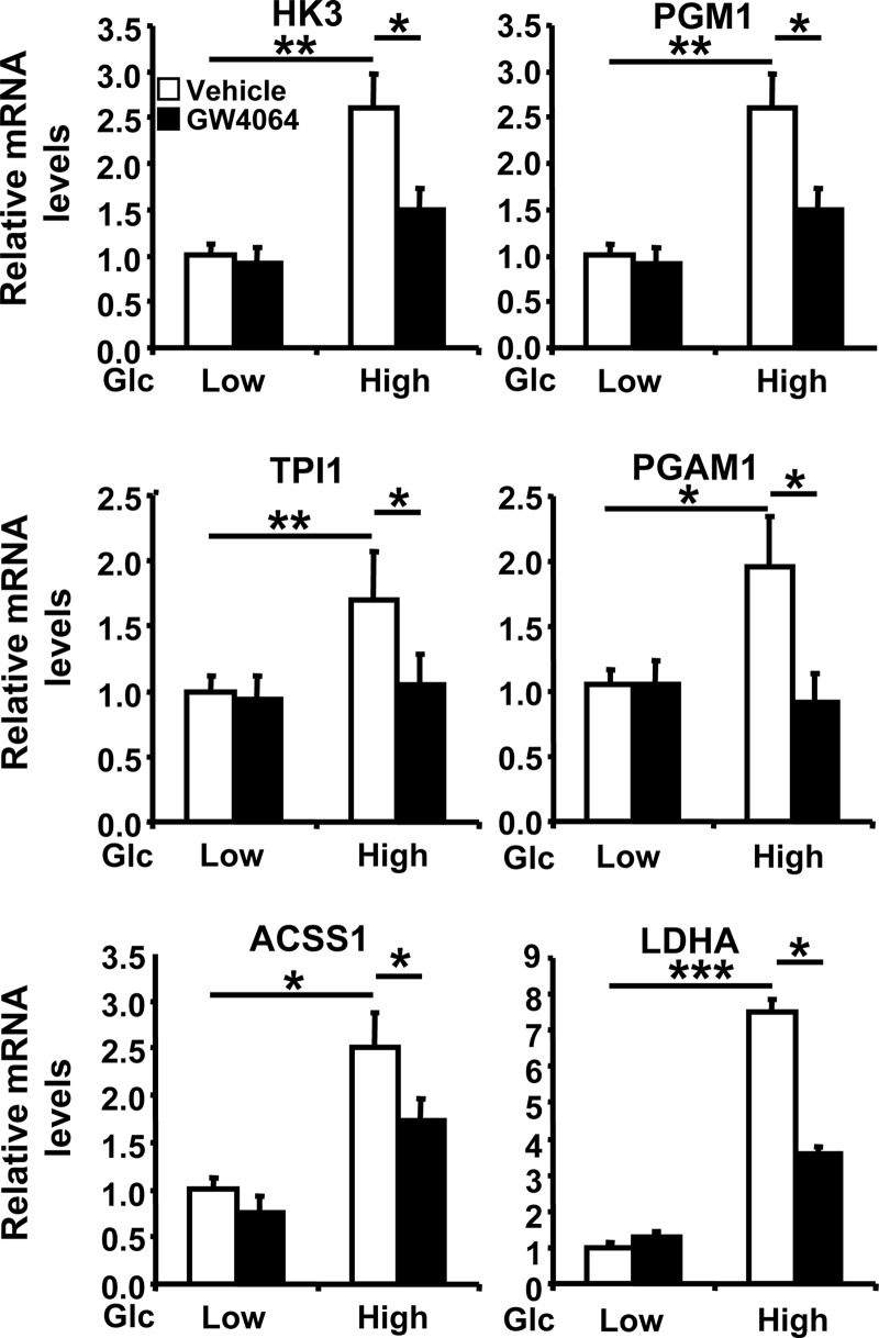
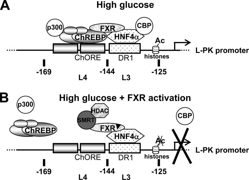
The glucose-activated transcription factor carbohydrate response element binding protein (ChREBP) induces the expression of hepatic glycolytic and lipogenic genes. The farnesoid X receptor (FXR) is a nuclear bile acid receptor controlling bile acid, lipid, and glucose homeostasis. FXR negatively regulates hepatic glycolysis and lipogenesis in mouse liver. The aim of this study was to determine whether FXR regulates the transcriptional activity of ChREBP in human hepatocytes and to unravel the underlying molecular mechanisms. Agonist-activated FXR inhibits glucose-induced transcription of several glycolytic genes, including the liver-type pyruvate kinase gene (L-PK), in the immortalized human hepatocyte (IHH) and HepaRG cell lines. This inhibition requires the L4L3 region of the L-PK promoter, known to bind the transcription factors ChREBP and hepatocyte nuclear factor 4α (HNF4α). FXR interacts directly with ChREBP and HNF4α proteins. Analysis of the protein complex bound to the L4L3 region reveals the presence of ChREBP, HNF4α, FXR, and the transcriptional coactivators p300 and CBP at high glucose concentrations. FXR activation does not affect either FXR or HNF4α binding to the L4L3 region but does result in the concomitant release of ChREBP, p300, and CBP and in the recruitment of the transcriptional corepressor SMRT. Thus, FXR transrepresses the expression of genes involved in glycolysis in human hepatocytes.


Figures

Fig 1
Fig 1
FXR activation inhibits the glucose-induced expression of the L-PK gene in mouse liver and IHH cells. (A) Glycemia (left) and hepatic L-PK mRNA levels (right) in wild-type mice subjected to 24 h of fasting (F) and then refed (RF) for 8 h with a high-carbohydrate diet after being pretreated (30 min) by gavage with vehicle alone (0.5% CMC–0.1% Tween 80) or INT-747 (30 mg/kg). Liver mRNA levels were measured by real-time quantitative PCR. Values are expressed relative to those in the fasting state, arbitrarily set to 1. (B) L-PK mRNA expression in IHH incubated for 24 h in a medium containing low (1 mM) or high (11 mM) glucose concentrations and vehicle (DMSO) or GW4064 (5 μM), WAY362450 (5 μM), INT-747 (10 μM), or chenodeoxycholic acid (CDCA, 100 μM). (C) Effects of FXR silencing on L-PK mRNA (top) and FXR protein (bottom) levels in IHH incubated for 24 h at low (1 mM) or high (11 mM) glucose concentrations and with vehicle (DMSO) or GW4064 (5 μM). L-PK and control 36B4 mRNA levels were measured by real-time quantitative PCR, and values are expressed relative to those at low glucose concentration with vehicle, arbitrarily set to 1. Total protein was extracted and analyzed as indicated in Materials and Methods. FXR protein levels were quantified by densitometry and normalized to the actin protein level. (D) Schematic representation of the L4L3 (−169 to −125) region of the human L-PK promoter that contains the ChREBP (ChORE) and HNF4α (DR-1) binding sites. (E) Activity of the L4L3 region of the L-PK promoter in IHH cells transfected with pGL3-TK-(L4L3)-LPK and incubated for 24 h in medium containing low (1 mM) or high (11 mM) glucose concentrations and vehicle (DMSO) or GW4064 (5 μM). Values are normalized to β-galactosidase activity and are expressed relative to those of cultures with pGL3-TK-(L4L3)-LPK and pCMV-Sport6 (B), pcDNA3 (C), or pGL3-TK-(L4L3)-LPK with vehicle at low glucose concentration (D), which were arbitrarily set to 1.
Fig 2
Fig 2
FXR physically interacts with ChREBP and HNF4α in vitro. (A) ChREBP protein levels in nuclear extracts from IHH incubated for 24 h in a medium containing low (1 mM) or high (11 mM) glucose concentrations and vehicle (DMSO) or GW4064 (5 μM). The expression of ChREBP protein was analyzed by Western blotting using a specific antibody. (B) In vitro GST pulldown experiments using full-length GST-FXR and in vitro transcribed/translated (TNT) ChREBP, Mlx, or HNF4α in the presence of [35S]methionine. (C) In vitro GST pulldown experiments using GST (lane 2) and the indicated FXR deletion mutant protein (lanes 3 and 4) and TNT ChREBP in the presence of [35S]methionine and vehicle (DMSO) or FXR ligand (GW4064, 5 μM). AF-1, activation function 1; DBD, DNA binding domain; LBD, ligand binding domain. (D) Two distinct anti-FXR antibodies were used for coimmunoprecipitation of ChREBP in nuclear extracts from IHH cells transfected with pSG5-FXR, pCMV-Sport6-ChREBP, and pcDNA3-Mlx and incubated in a medium containing a high (11 mM) glucose concentration. The expression of FXR and ChREBP proteins was detected 24 h after transfection by Western blotting (WB) using specific antibodies (AB1 and AB2).
Fig 3
Fig 3
FXR activation at high glucose concentrations releases ChREBP but not FXR and HNF4α from the L4L3 region of the L-PK promoter. Relative levels of L-PK promoter occupancy by ChREBP, HNF4α, and FXR on the L4L3 region are shown. The levels of occupancy were evaluated by quantitative PCR in ChIP experiments performed using total extracts from IHH incubated for 5 h in medium containing a low (1 mM) or high (11 mM) glucose concentration and vehicle (DMSO) or GW4064 (5 μM). Occupancies are expressed relative to those at low glucose concentration with vehicle, arbitrarily set to 1. Each experiment was performed at least 3 times, and results are the averages and standard deviations of these experiments.
Fig 4
Fig 4
FXR activation leads to the release of coactivators p300 and CBP and the recruitment of the coinhibitor SMRT on the L4L3 region at high glucose concentration. (A) Relative levels of L-PK promoter occupancy by p300 and CBP on the L4L3 region. (B) Effects of SMRT gene silencing on L-PK mRNA (left) and SMRT protein (right) levels in IHH transfected with specific siRNAs and incubated for 24 h at low (1 mM) or high (11 mM) glucose concentration and with vehicle (DMSO) or GW4064 (5 μM). Proteins were extracted and analyzed as indicated in Materials and Methods. SMRT protein levels were quantified by densitometry and normalized to actin protein level. (C) In vitro GST pulldown experiments using full-length GST-SMRT and TNT FXR in the presence of [35S]methionine. (D) Relative levels of L-PK promoter occupancy by SMRT. (E) Effects of TSA treatment on L-PK gene expression in IHH incubated for 24 h at low (1 mM) or high (11 mM) glucose concentration and with vehicle (DMSO) or GW4064 (5 μM). (F) Relative levels of H3K9 acetylation of L-PK. For the experiments whose results are shown in panels A, D, and F, the occupancies were evaluated by quantitative PCR in ChIP experiments performed using total extracts from IHH incubated for 5 h in a medium containing low (1 mM) or high (11 mM) glucose concentrations and vehicle (DMSO) or GW4064 (5 μM). Occupancies are expressed relative to those at low glucose concentration with vehicle, arbitrarily set to 1. Each experiment was performed at least 3 times, and the results are the averages and standard deviations of these experiments. For the experiments whose results are shown in panels B (left) and E, L-PK and control 36B4 mRNA levels were measured by real-time quantitative PCR. The values are expressed relative to those at low glucose concentration with vehicle, which were arbitrarily set to 1.
Fig 5
Fig 5
The expression of other ChREBP-regulated genes is also inhibited by FXR by involving a molecular mechanism similar to that for L-PK. (A) mRNA expression of ChREBP target genes in IHH incubated for 24 h in a medium containing low (1 mM) or high (11 mM) glucose concentrations and vehicle (DMSO) or GW4064 (5 μM). Gene and control 36B4 mRNA levels were measured by real-time quantitative PCR. Values are expressed relative to those measured at low glucose concentration with vehicle, arbitrarily set to 1. (B) Relative levels of TxNIP promoter occupancy by ChREBP and FXR (top), p300 and CBP (middle), and SMRT and H3K9 (bottom). The occupancies were evaluated by quantitative PCR amplification of the region of the TxNIP promoter that contains the ChORE in ChIP experiments performed using total extracts from IHH incubated for 5 h at low (1 mM) or high (11 mM) glucose concentrations and vehicle (DMSO) or GW4064 (5 μM). Occupancies are expressed relative to those at low glucose concentration with vehicle, arbitrarily set to 1. Each experiment was performed at least three times, and results from a representative experiment are shown.
Fig 6
Fig 6
FXR inhibits the expression of glucose-induced genes. Genes identified as being regulated by both glucose and FXR using microarray analysis were analyzed for mRNA expression in IHH incubated for 24 h in a medium containing low (1 mM) or high (11 mM) glucose concentrations and vehicle (DMSO) or GW4064 (5 μM). HK3, hexokinase 3; PGM1, phosphoglucomutase 1; TPI1, triosephosphate isomerase 1; PGAM1, phosphoglycerate mutase 1; ACSS1, acyl-CoA synthetase short-chain family member 1; LDHA, lactate dehydrogenase A. Gene and control 36B4 mRNA levels were measured by real-time quantitative PCR. The values are expressed relative to those at low glucose concentration with vehicle, which were arbitrarily set to 1.
Fig 7
Fig 7
Model of transrepression of glucose-induced L-PK gene expression by FXR. (A) At high glucose concentrations without FXR activation, ChREBP and HNF4α are bound on the L4L3 region of the L-PK promoter and trans activate gene expression, in part due to the recruitment of the transcriptional coactivators p300 and CBP. FXR is integrated into this protein complex, probably through its direct interaction with ChREBP and HNF4α. (B) At high glucose concentration after FXR activation, ChREBP, as well as p300 and CBP, is released from the L-PK promoter. FXR and HNF4α are still bound on the L-PK promoter. Tethered to the promoter through its interaction with HNF4α, FXR recruits transcriptional coinhibitor SMRT and represses the transcription through the recruitment of HDACs and deacetylation of H3 histones.

References

    1. Bouché C, Serdy S, Kahn CR, Goldfine AB. 2004. The cellular fate of glucose and its relevance in type 2 diabetes. Endocr. Rev. 25:807–830 - PubMed
    1. Nordlie RC, Foster JD, Lange AJ. 1999. Regulation of glucose production by the liver. Annu. Rev. Nutr. 19:379–406 - PubMed
    1. Girard J, Ferré P, Foufelle F. 1997. Mechanisms by which carbohydrates regulate expression of genes for glycolytic and lipogenic enzymes. Annu. Rev. Nutr. 17:325–352 - PubMed
    1. de Luis O, Valero MC, Jurado LA. 2000. WBSCR14, a putative transcription factor gene deleted in Williams-Beuren syndrome: complete characterisation of the human gene and the mouse ortholog. Eur. J. Hum. Genet. 8:215–222 - PubMed
    1. Yamashita H, Takenoshita M, Sakurai M, Bruick RK, Henzel WJ, Shillinglaw W, Arnot D, Uyeda K. 2001. A glucose-responsive transcription factor that regulates carbohydrate metabolism in the liver. Proc. Natl. Acad. Sci. U. S. A. 98:9116–9121 - PMC - PubMed

Publication types

MeSH terms