. 2013 Apr 8;210(4):655-63.
doi: 10.1084/jem.20122824. Epub 2013 Mar 25.

Engineering HIV envelope protein to activate germline B cell receptors of broadly neutralizing anti-CD4 binding site antibodies

Affiliations

Engineering HIV envelope protein to activate germline B cell receptors of broadly neutralizing anti-CD4 binding site antibodies

Andrew T McGuire et al. J Exp Med. .

Abstract

Broadly neutralizing antibodies (bnAbs) against HIV are believed to be a critical component of the protective responses elicited by an effective HIV vaccine. Neutralizing antibodies against the evolutionarily conserved CD4-binding site (CD4-BS) on the HIV envelope glycoprotein (Env) are capable of inhibiting infection of diverse HIV strains, and have been isolated from HIV-infected individuals. Despite the presence of anti-CD4-BS broadly neutralizing antibody (bnAb) epitopes on recombinant Env, Env immunization has so far failed to elicit such antibodies. Here, we show that Env immunogens fail to engage the germline-reverted forms of known bnAbs that target the CD4-BS. However, we found that the elimination of a conserved glycosylation site located in Loop D and two glycosylation sites located in variable region 5 of Env allows Env-binding to, and activation of, B cells expressing the germline-reverted BCRs of two potent broadly neutralizing antibodies, VRC01 and NIH45-46. Our results offer a possible explanation as to why Env immunogens have been ineffective in stimulating the production of such bNAbs. Importantly, they provide key information as to how such immunogens can be engineered to initiate the process of antibody-affinity maturation against one of the most conserved Env regions.


Figures

Figure 1.
Figure 1.
Env binding to the mature and germline-reverted NIH45-46 BCRs and B cell activation. (a) NIH45-46 IgG binding to the indicated recombinant trimeric gp140 Envs was measured by ELISA. (b) B cells expressing the mature (white bars) or germline-reverted (gray bars) NIH45-46 BCR, as well as mock transfected cells (black bars), were incubated with the indicated trimeric gp140 proteins, followed by incubation with a PE-labeled polyclonal anti-HIV IgG pool. The MFI of PE staining was determined in NIH45-46 BCR expressing cells identified by staining with APC-labeled monoclonal anti–human IgG. Error bars represent SD from the mean of two independent experiments. (c and d) B cells expressing the mature (c) or germline-reverted (d) NIH45-46 BCR were loaded with Fluo-4 direct calcium indicator and stimulated with the indicated trimeric gp140 proteins. Black arrows in c and d indicate time of Env-addition. Data are representative of three independent experiments.
Figure 2.
Figure 2.
Removal of N-linked glycosylation sites from the 426c Env confers binding to germline-reverted NIH45-46 and VRC01. Binding of mature NIH45-46 (a, d, g, and j), germline-reverted NIH45-46 (b, e, h, and k), and germline-reverted VRC01(c, f, i, and l) IgG to trimeric gp140 426c (a–c; wild type), 426c lacking the NLGS at position 276 (d–f; N276D), 426c lacking NLGS at positions 460 and 463 (g–i; N460D.N463D), or 426c lacking NLGS at positions 276, 460, and 463 (j–l; N276D.N460D.N463D) was measured by BLI. For mature and germline-reverted NIH45-46, black, red, orange, blue, and green lines represent 1.7 µM, 850 nM, 425 nM, 212 nM, and 106 nM of trimeric Env, respectively. For germline-reverted VRC01, black, red, orange, blue, and green lines represent 2 µM, 1 µM, 500 nM, 250 nM, and 125 nM of trimeric Env, respectively. Dashed gray lines represent the theoretical fit. The binding kinetic data (including experimental error) are summarized in Table S3.
Figure 3.
Figure 3.
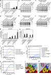
Removal of N-linked glycosylation sites from the 426c Env results in stimulation of the germline-reverted NIH45-46 and VRC01 BCRs. (a) B cells expressing the mature (white bars) or germline-reverted (black bars) NIH45-46 IgG BCR, were incubated with the indicated trimeric gp140 proteins, followed by incubation with a PE-labeled polyclonal anti-HIV IgG pool. The MFI of PE staining was determined in NIH45-46 BCR expressing cells identified by staining with APC-labeled monoclonal anti–human IgG. Error bars represent SD from the mean of two independent experiments (b–f). B cells expressing the mature (b), germline-reverted NIH45-46 (c), and germline-reverted VRC01 (d) IgG BCR, as well as the germline-reverted NIH45-46 (e), and germline-reverted VRC01 (f) IgM BCR were either unstimulated, or were stimulated with the indicated 426c Env variants (40 µg/ml for the mature BCR, or 200 µg/ml for the germline-reverted BCRs). Lysates were separated by SDS-PAGE, transferred to nitrocellulose, and stained with the mouse anti-phosphotyrosine mAb 4G10 (top) and a rabbit anti–β-actin mAb 13E5 (bottom), followed by IRDye 800CW-labeled goat anti–mouse and IRDye 680LT-labeled goat anti–rabbit antibodies. Black arrows indicate proteins that show an increase in tyrosine phosphorylation. (g) Quantification of tyrosine phosphorylation signal corresponding to protein band in b–d are denoted with #. White bars, mature NIH45-46; black bars, germline NIH45-46; gray bars, germline VRC01. (h) Quantification of tyrosine phosphorylation signal corresponding to protein band in e and f, denoted with arrow. Black bars (germline NIH45-46) and gray bars (germline VRC01). Signal intensity was normalized to the actin loading controls, and expressed relative to the unstimulated cells. Error bars represent the SD from two (germline-reverted IgG and IgM VRC01) or three (mature and germline-reverted IgG and IgM NIH45-46) independent experiments. *, actin-normalized log-transformed signal intensities that are statistically different from the unstimulated controls using a repeated measures ANOVA methodology, followed by Tukey’s multiple comparisons test. (i–k) B cells expressing the mature NIH45-46 (i), germline-reverted NIH45-46 (j), or germline-reverted VRC01 (k) IgG BCR were loaded with Fluo-4 direct calcium indicator and stimulated with the indicated Env proteins. Black arrow indicates time of Env addition. (l) Model illustrating how NIH45-46 (transparent) clashes with glycans at gp120 residues 276 and 460 if glycans are conformationally relaxed in the absence of NIH45-46. (m) Model of NIH45-46 bound to gp120 with glycans removed from 276, 460, and 463 to eliminate potential antibody-glycan clash. Red, gp120; yellow, NIH45-46 contact residues on gp120; blue, NIH45-46 heavy chain; purple, NIH45-46 light chain; orange, glycan at 276; green, glycan at 460; cyan, glycan at 463; white, glycans at other positions.
Figure 4.
Figure 4.
Neutralization of 426c viral variants by NIH45-46. (a and b)Entry of pseudovirions expressing either the wild-type 426c Env or derivatives lacking NLGS at positions 276, 460, and 463 into TZM-bl cells was measured in the presence of mature (a) or germline-reverted (b) NIH45-46 IgG. Pseudovirions expressing the unrelated murine leukemia virus (MLV) envelope are included as controls. Entry of the indicated pseudovirions into TZM-bl cells in the presence of mAb PG9 was used as control for nonspecific neutralization that may sometimes occur at high mAb concentrations (c). Each data point represents the mean ± SEM of relative luciferase units of two independent experiments, each done in duplicate. (d) Single-round entry competent pseudovirions expressing the indicated 426c Env were added to TZMbl cells in a 96-well microtiter plate at 0.1 ng/ well p24 (n = 6; open bars), and 1 ng/ well p24 (n = 6; filled bars). Bars represent the mean ± SEM of relative luciferase units expressed relative to that obtained with the wild-type virus (n = 6).
Figure 5.
Figure 5.
Binding of mature and germline-reverted anti–CD4-BS bnmAbs to the 426 Env lacking NLGS at positions 276, 460, and 463. Binding of the mature (a) and germline-reverted (b) antibodies to 426c Env lacking all three NLGS at a concentration of 1 µM was measured by BLI. Binding of the germline-reverted NIH45-46 light chain with the germline-reverted 3BNC60/3BNC117 heavy chain (c) and the germline-reverted NIH45-46 light chain with the germline-reverted 12A21 heavy chain (d) to the indicated trimeric Env was measured by BLI. All Env were tested at concentration of 1 µM.

References

    1. Binley J.M., Lybarger E.A., Crooks E.T., Seaman M.S., Gray E., Davis K.L., Decker J.M., Wycuff D., Harris L., Hawkins N., et al. 2008. Profiling the specificity of neutralizing antibodies in a large panel of plasmas from patients chronically infected with human immunodeficiency virus type 1 subtypes B and C. J. Virol. 82:11651–11668 10.1128/JVI.01762-08 - DOI - PMC - PubMed
    1. Diskin R., Scheid J.F., Marcovecchio P.M., West A.P., Jr, Klein F., Gao H., Gnanapragasam P.N., Abadir A., Seaman M.S., Nussenzweig M.C., Bjorkman P.J. 2011. Increasing the potency and breadth of an HIV antibody by using structure-based rational design. Science. 334:1289–1293 10.1126/science.1213782 - DOI - PMC - PubMed
    1. Gray E.S., Madiga M.C., Hermanus T., Moore P.L., Wibmer C.K., Tumba N.L., Werner L., Mlisana K., Sibeko S., Williamson C., et al. ; CAPRISA002 Study Team 2011a. The neutralization breadth of HIV-1 develops incrementally over four years and is associated with CD4+ T cell decline and high viral load during acute infection. J. Virol. 85:4828–4840 10.1128/JVI.00198-11 - DOI - PMC - PubMed
    1. Gray G.E., Allen M., Moodie Z., Churchyard G., Bekker L.G., Nchabeleng M., Mlisana K., Metch B., de Bruyn G., Latka M.H., et al. ; HVTN 503/Phambili study team 2011b. Safety and efficacy of the HVTN 503/Phambili study of a clade-B-based HIV-1 vaccine in South Africa: a double-blind, randomised, placebo-controlled test-of-concept phase 2b study. Lancet Infect. Dis. 11:507–515 10.1016/S1473-3099(11)70098-6 - DOI - PMC - PubMed
    1. Hoot S., McGuire A.T., Cohen K.W., Strong R.K., Hangartner L., Klein F., Diskin R., Scheid J.F., Sather D.N., Burton D.R., Stamatatos L. 2013. Recombinant HIV envelope proteins fail to engage germline versions of anti-CD4bs bNAbs. PLoS Pathog. 9:e1003106 10.1371/journal.ppat.1003106 - DOI - PMC - PubMed

Publication types

MeSH terms