Comparative Study
. 2013 Apr 16;110(16):E1462-9.
doi: 10.1073/pnas.1216894110. Epub 2013 Mar 25.

Genomic assay reveals tolerance of DNA damage by both translesion DNA synthesis and homology-dependent repair in mammalian cells

Affiliations
Comparative Study

Genomic assay reveals tolerance of DNA damage by both translesion DNA synthesis and homology-dependent repair in mammalian cells

Lior Izhar et al. Proc Natl Acad Sci U S A. .

Abstract

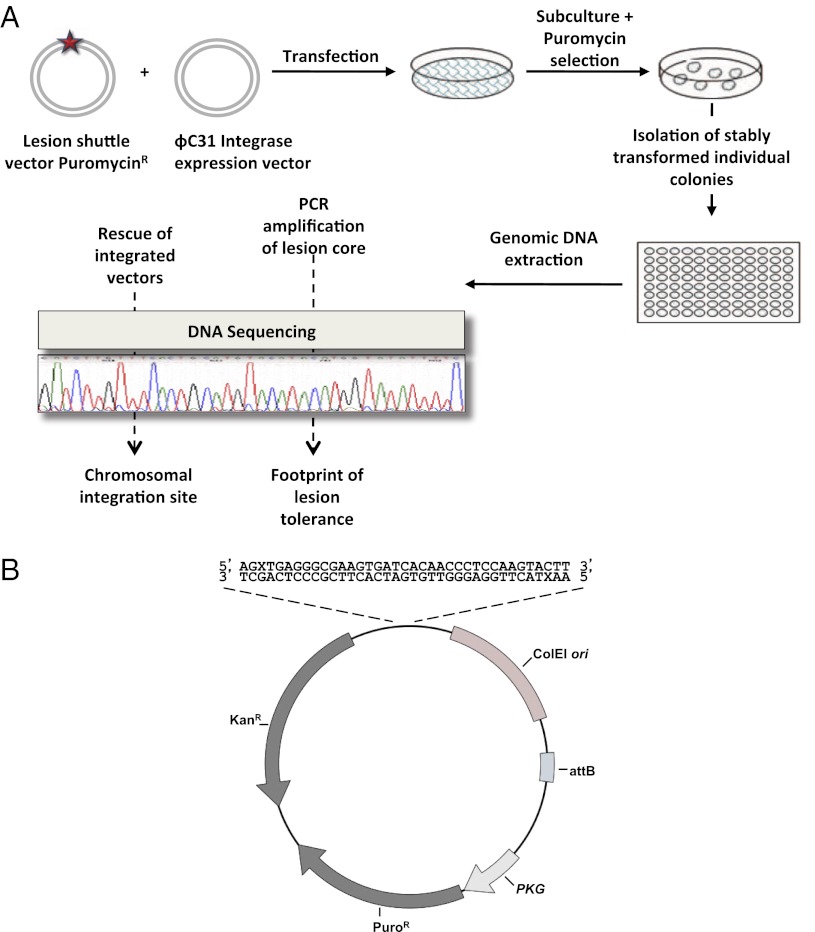
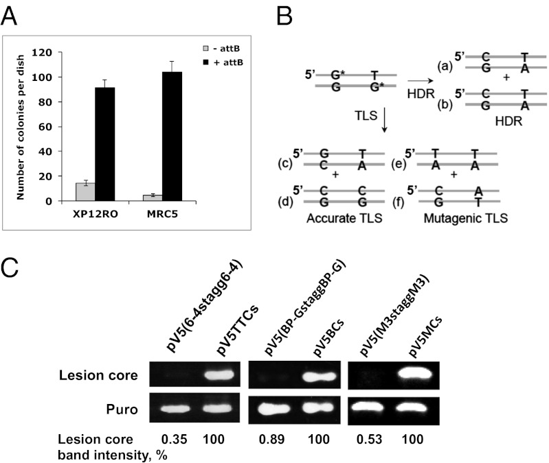
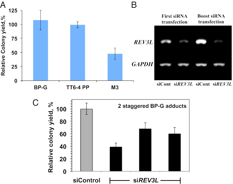
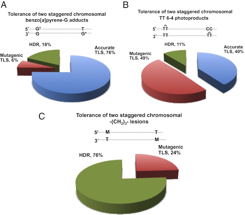
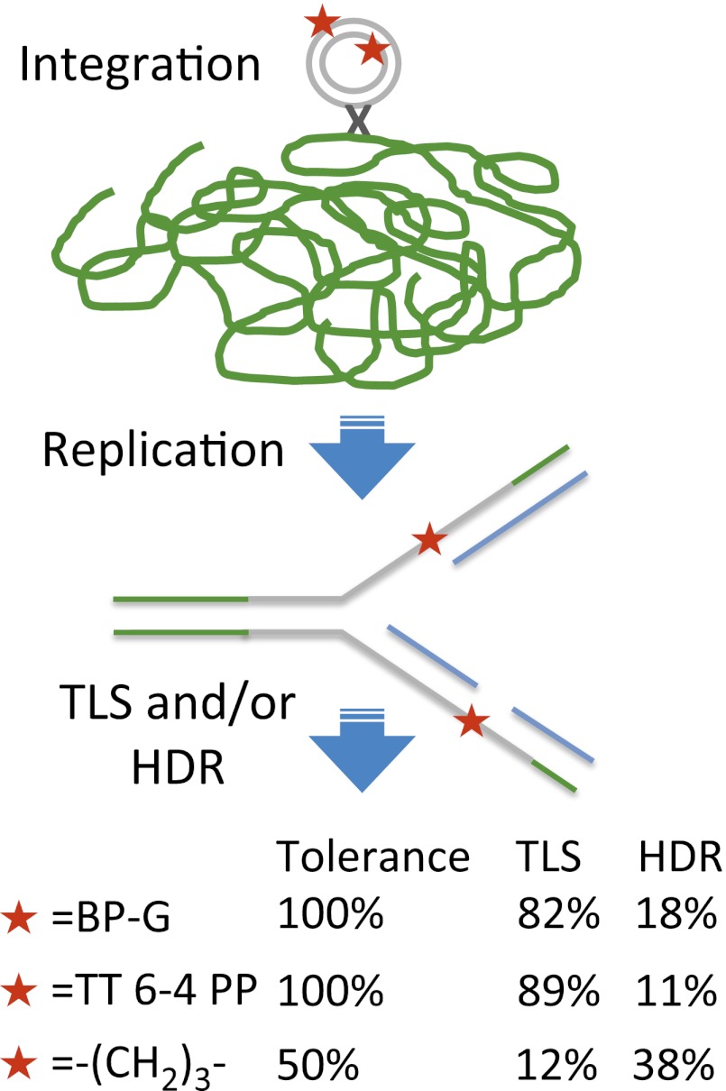
DNA lesions can block replication forks and lead to the formation of single-stranded gaps. These replication complications are mitigated by DNA damage tolerance mechanisms, which prevent deleterious outcomes such as cell death, genomic instability, and carcinogenesis. The two main tolerance strategies are translesion DNA synthesis (TLS), in which low-fidelity DNA polymerases bypass the blocking lesion, and homology-dependent repair (HDR; postreplication repair), which is based on the homologous sister chromatid. Here we describe a unique high-resolution method for the simultaneous analysis of TLS and HDR across defined DNA lesions in mammalian genomes. The method is based on insertion of plasmids carrying defined site-specific DNA lesions into mammalian chromosomes, using phage integrase-mediated integration. Using this method we show that mammalian cells use HDR to tolerate DNA damage in their genome. Moreover, analysis of the tolerance of the UV light-induced 6-4 photoproduct, the tobacco smoke-induced benzo[a]pyrene-guanine adduct, and an artificial trimethylene insert shows that each of these three lesions is tolerated by both TLS and HDR. We also determined the specificity of nucleotide insertion opposite these lesions during TLS in human genomes. This unique method will be useful in elucidating the mechanism of DNA damage tolerance in mammalian chromosomes and their connection to pathological processes such as carcinogenesis.


Conflict of interest statement

The authors declare no conflict of interest.

Figures

Fig. 1.
Fig. 1.
Principle of the DNA damage tolerance assay for chromosomes of mammalian cells. (A) Schematic presentation of the two cellular strategies for DNA damage tolerance, TLS and HDR. (B) Principle of the unique TLS/HDR method, based on phage integrase-mediated integration into mammalian chromosomes of a plasmid carrying a site-specific lesion. See text for details.
Fig. 2.
Fig. 2.
Outline of the DNA damage tolerance assay in chromosomes of mammalian cells. (A) Cultured cells were cotransfected with the lesion shuttle vector and the ϕC31 expression plasmid. In parallel, cells were transfected with a similarly constructed control plasmid without a lesion. Forty-eight hours after transfection the cells were subcultured, and after 24 additional hours they were subjected to puromycin selection. Approximately 14 d later resistant colonies were counted, picked, and individually transferred into a 96-well plate for further growth. When they reached suitable confluence, the cells in each individual well were harvested, and their chromosomal DNA was extracted. This DNA was used as a template in PCR reactions aimed to amplify the lesion core area. Amplified samples were then subjected to DNA sequence analysis. (B) Structure of the lesion shuttle vector pLSV5L. PuroR, puromycin resistance gene under the phosphoglycerate kinase (PGK) promoter; attB, phage ϕC31 integrase attachment site; kanR, kanamycin resistance gene; X stands for a lesion. Two lesions in the staggered configuration are shown.
Fig. 3.
Fig. 3.
(A) Effect of ϕC31-integrase activity on the efficiency of plasmid integration into the chromosomes of mammalian cells. Human XP12RO and MRC5 cells were cotransfected with a mixture of the integrase expression vector pKGϕC31-int and pLSV5 (black bar) or pLSV5nA (gray bar) that lacks the integrase recognition sequence attB. Following transfection the cells were grown under puromycin selection for 12 d, until visible colonies were formed. Culture plates were then fixed and counted. The results shown are an average of three different transfections. (B) Products expected from TLS and HDR of two staggered BP-G lesions during chromosomal DNA damage tolerance. (C) Persistence of DNA lesions in XPA cells. Human XP12RO cells were transfected with the lesion shuttle vectors pLSV5TT6-4stagg6-4, pLSV5BP-GstaggBP-G, pLSV5M3staggM3, or their corresponding control vectors (without lesions). The vectors were extracted from the cells 3 d after transfection and used as templates in PCR reactions aimed to amplify the region containing the lesions (lesion core) and in parallel PCR reactions aimed to amplify a fragment of the puromycin resistance gene (puro) as an internal reference. Quantification of the images of the ethidium bromide-stained gel was done using ImageJ software. The percentage of the lesion-free background in each lesion shuttle vector is shown relative to the corresponding control vector with no lesion (100%), normalized to the intensity of the “puro” bands for each plasmid.
Fig. 4.
Fig. 4.
Analysis of the tolerance of DNA lesions integrated into human chromosomes. (A) Relative colony yields of tolerance of two-staggered DNA lesions integrated into the chromosomes of human XPA cells. The detailed data are presented in Tables S1S6. (B) RT-PCR quantification of REV3L mRNA after treatment with siRNA and a boost treatment after 24 additional hours. (C) Relative colony yields of tolerance of two-staggered BP-G adducts in XPA cells treated with REV3L siRNA or a control siRNA. Results of three different transfections are presented.
Fig. 5.
Fig. 5.
Categories of DNA damage tolerance events of DNA lesions integrated into human chromosomes. (A–C) The data shown are for pairs of BP-G adducts (A), TT 6–4 photoproducts (B), and M3 lesions (C), in a staggered configuration. The detailed data are presented in Tables S1S6, respectively.
Fig. P1.
Fig. P1.
Assay for the simultaneous measurement of TLS and HDR across defined lesions in mammalian cultured cells. A portable DNA vector carrying defined lesions was integrated into mammalian chromosomes, using a bacteriophage-encoded integrase. The vector carried defined lesions on each strand, to ensure that once integrated, each strand must undergo DNA damage tolerance to complete replication. The nucleotides opposite the lesions were engineered to allow distinction between TLS and HDR. Tolerance of a tobacco smoke-induced DNA lesion (termed BP-G) and a UV light-induced DNA lesion (termed TT 6–4 PP) occurred with an efficiency of about 100%, allowing survival of the cells. In contrast, tolerance of an artificial trimethylene segment occurred in only half of the cells, whereas the other half died. DNA sequence analysis indicated that all three lesions were bypassed by both TLS and HDR. TLS across BP-G was largely accurate, but highly mutagenic across TT 6–4 PP.

References

    1. Friedberg EC, et al. DNA Repair and Mutagenesis. 2nd Ed. Washington, DC: ASM Press; 2006.
    1. Friedberg EC. Suffering in silence: The tolerance of DNA damage. Nat Rev Mol Cell Biol. 2005;6(12):943–953. - PubMed
    1. Lehmann AR, Fuchs RP. Gaps and forks in DNA replication: Rediscovering old models. DNA Repair. 2006;5(12):1495–1498. - PubMed
    1. Prakash S, Johnson RE, Prakash L. Eukaryotic translesion synthesis DNA polymerases: Specificity of structure and function. Annu Rev Biochem. 2005;74:317–353. - PubMed
    1. Livneh Z, Ziv O, Shachar S. Multiple two-polymerase mechanisms in mammalian translesion DNA synthesis. Cell Cycle. 2010;9(4):729–735. - PubMed

Publication types

LinkOut - more resources