. 2013 Apr 9;110(15):5875-80.
doi: 10.1073/pnas.1222419110. Epub 2013 Mar 25.

Characterization of platelet aminophospholipid externalization reveals fatty acids as molecular determinants that regulate coagulation

Affiliations

Characterization of platelet aminophospholipid externalization reveals fatty acids as molecular determinants that regulate coagulation

Stephen R Clark et al. Proc Natl Acad Sci U S A. .

Abstract

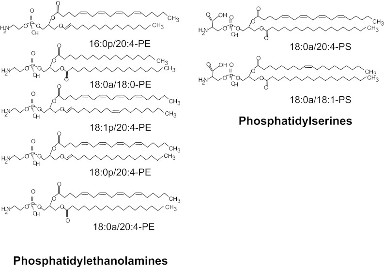
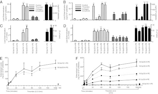
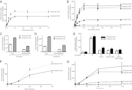
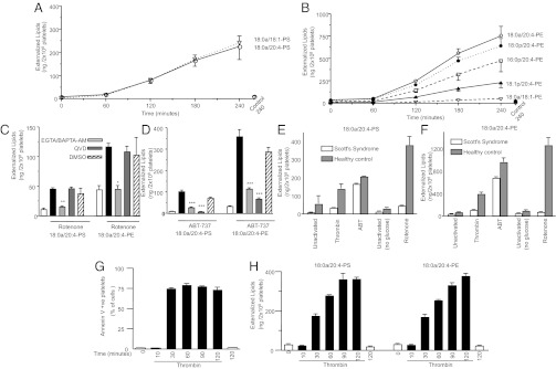
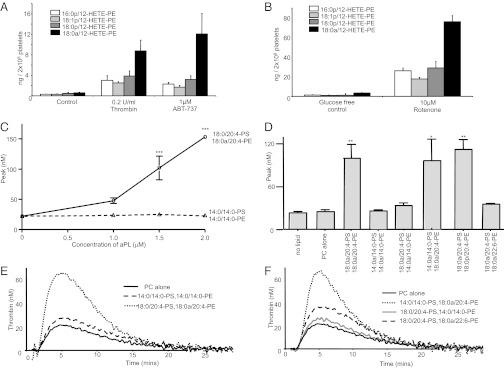
Aminophospholipid (APL) trafficking across the plasma membrane is a key event in cell activation, apoptosis, and aging and is required for clearance of dying cells and coagulation. Currently the phospholipid molecular species externalized are unknown. Using a lipidomic method, we show that thrombin, collagen, or ionophore-activated human platelets externalize two phosphatidylserines (PSs) and five phosphatidylethanolamines (PEs). Four percent of the total cellular PE/PS pool (∼300 ng/2 × 10(8) cells, thrombin), is externalized via calcium mobilization and protease-activated receptors-1 and -4, and 48% is contained in microparticles. Apoptosis and energy depletion (aging) externalized the same APLs in a calcium-dependent manner, and all stimuli externalized oxidized phospholipids, termed hydroxyeicosatetraenoic acid-PEs. Transmembrane protein-16F (TMEM-16F), the protein mutated in Scott syndrome, was required for PE/PS externalization during thrombin activation and energy depletion, but not apoptosis. Platelet-specific APLs optimally supported tissue factor-dependent coagulation in human plasma, vs. APL with longer or shorter fatty acyl chains. This finding demonstrates fatty acids as molecular determinants of APL that regulate hemostasis. Thus, the molecular species of externalized APL during platelet activation, apoptosis, and energy depletion were characterized, and their ability to support coagulation revealed. The findings have therapeutic implications for bleeding disorders and transfusion therapy. The assay could be applied to other cell events characterized by APL externalization, including cell division and vesiculation.


Conflict of interest statement

The authors declare no conflict of interest.

Figures

Fig. 1.
Fig. 1.
Structures of APL externalized by human platelets.
Fig. 2.
Fig. 2.
Several APL molecular species are externalized by human platelets in response to agonists and are dose-dependent to thrombin. (A and B) Platelets were activated using thrombin (0.2 U/mL), collagen (10 µg/mL), or ionophore (A23187, 10 µM) at 37° for 30 min, before biotinylation, lipid extraction, and LC/MS/MS analysis as in Materials and Methods. (C and D) Total APL in each sample was determined by biotinylation using NB, as described in Materials and Methods, and used to calculate the relative proportion externalized on platelet activation. (E and F) Platelets were activated by thrombin (0.2 U/mL) for varying times, as shown, before biotinylation, lipid extraction, and LC/MS/MS analysis as in Materials and Methods (n = 3; mean ± SEM; data representative of three independent experiments. *P < 0.05, **P < 0.01, and ***P < 0.001 vs. control, using ANOVA and Bonferroni post hoc test).
Fig. 3.
Fig. 3.
Externalization of APL is time dependent, associated with microparticles, requires calcium, and occurs during apoptosis. (A and B) Platelets were activated with varying amounts of thrombin for 120 min, before biotinylation, lipid extraction, and LC/MS/MS analysis as in Materials and Methods. (C and D) Platelets were activated using thrombin (0.2 U/mL, 30 min), then centrifuged as described in Materials and Methods. Supernatant (microparticles) and pellets were separately analyzed for surface exposed APLs. (E) Platelets were activated using thrombin (0.2 U/mL, 30 min) after 10 min preincubation with Ca2+ chelators (EGTA, 1mM and/or BAPTA-AM, 10 μM). (F and G) Platelets were activated using 1 μM ABT-737 for varying times before lipid extraction and analysis. For all experiments, n = 3; mean ± SEM; data representative of three independent donors. *P < 0.05, **P < 0.01, and ***P < 0.001 vs. control, using ANOVA and Bonferroni post hoc test.
Fig. 4.
Fig. 4.
(i) Energy depletion causes similar APL externalization to thrombin, (ii) energy depletion and apoptosis APL exposure require calcium mobilization, (iii) TMEM-16F is required for APL externalization in response to thrombin, or energy depletion, but not apoptosis, and (iv) annexin V-FITC binding vs. biotinylation of APL reveals different kinetics. (A and B) Platelets were activated using 10 μM rotenone for varying times before lipid extraction and analysis. (C and D) Platelets were activated with 10 μM rotenone for 180 min, or with 1 μM ABT-737 for 60 min, after preincubation with inhibitors (Q-VD-OPH, caspase inhibitor, 10 μM), EGTA (1 mM), BAPTA-AM (10 μM), then surface APL analyzed as described in Materials and Methods. (E and F) Scott syndrome or healthy control platelets were activated with 0.2 U/mL thrombin for 30 min, 1 μM ABT-737 for 60 min, or 10 μM rotenone (in the absence of glucose) for 180 min, then surface APL analyzed as described in Materials and Methods. (G and H) Annexin V-FITC binding was determined using flow cytometry, or APL exposure measured using biotinylation, as described in Materials and Methods, after activation using 0.2 U/mL thrombin. For all experiments, n = 3; mean ± SEM; data representative of three independent donors. *P < 0.05, **P < 0.01, and ***P < 0.001 vs. rotenone or thrombin (+DMSO) samples, as appropriate, using ANOVA and Bonferroni post hoc test.
Fig. 5.
Fig. 5.
Thrombin, apoptosis, and energy depletion cause externalization of HETE-PEs, and tissue factor-dependent thrombin generation requires specific fatty acids on PE, but not PS. (A and B) Externalized HETE-PEs were determined on platelets activated using thrombin (30 min), ABT-737 (60 min) or rotenone/glucose-free (3 h), before biotinylation and analysis as described in Materials and Methods. Biotinylated 18:0a/12-HETE-PE was used as primary standard for quantitation. (C) Tissue factor (1 pM)-dependent thrombin generation was determined in human plasma using liposomes containing 4 μM PLs (with APL varying from 0 to 2 μM) of varying fatty acid composition, as described in Materials and Methods. (D) Tissue factor-dependent thrombin generation was determined using liposomes containing 1.5 µM APL (in a total of 4 µM PL), as described in Materials and Methods. APL composition is varied according to the labels on the figure, but PS:PE is maintained at 1:2.7 throughout. Representative experiments are shown in E and F. For all experiments, n = 3; mean ± SEM; data from three independent experiments. ***P < 0.001, **P < 0.01, *P < 0.05 vs. PC alone control, using a two-way ANOVA and Bonferroni post hoc test.

References

    1. Daleke DL. Phospholipid flippases. J Biol Chem. 2007;282(2):821–825. - PubMed
    1. Ravichandran KS. Find-me and eat-me signals in apoptotic cell clearance: Progress and conundrums. J Exp Med. 2010;207(9):1807–1817. - PMC - PubMed
    1. Heemskerk JW, Bevers EM, Lindhout T. Platelet activation and blood coagulation. Thromb Haemost. 2002;88(2):186–193. - PubMed
    1. Zwaal RF, Comfurius P, Bevers EM. Surface exposure of phosphatidylserine in pathological cells. Cell Mol Life Sci. 2005;62(9):971–988. - PMC - PubMed
    1. Daleke DL. Regulation of transbilayer plasma membrane phospholipid asymmetry. J Lipid Res. 2003;44(2):233–242. - PubMed

Publication types

LinkOut - more resources