. 2013 Apr 9;110(15):5887-92.
doi: 10.1073/pnas.1217823110. Epub 2013 Mar 25.

Dimers of mitochondrial ATP synthase form the permeability transition pore

Affiliations

Dimers of mitochondrial ATP synthase form the permeability transition pore

Valentina Giorgio et al. Proc Natl Acad Sci U S A. .

Abstract

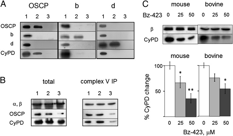
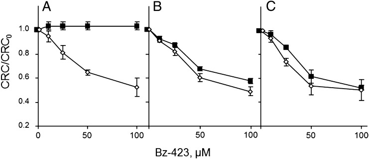
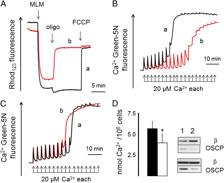
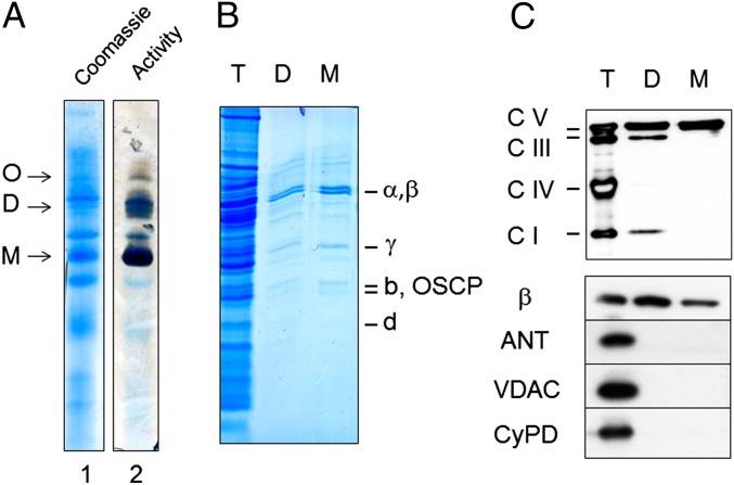
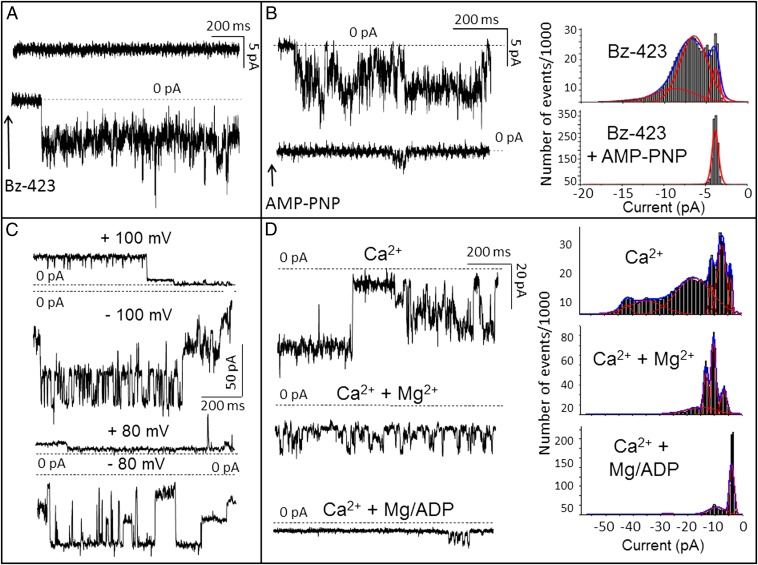
Here we define the molecular nature of the mitochondrial permeability transition pore (PTP), a key effector of cell death. The PTP is regulated by matrix cyclophilin D (CyPD), which also binds the lateral stalk of the FOF1 ATP synthase. We show that CyPD binds the oligomycin sensitivity-conferring protein subunit of the enzyme at the same site as the ATP synthase inhibitor benzodiazepine 423 (Bz-423), that Bz-423 sensitizes the PTP to Ca(2+) like CyPD itself, and that decreasing oligomycin sensitivity-conferring protein expression by RNAi increases the sensitivity of the PTP to Ca(2+). Purified dimers of the ATP synthase, which did not contain voltage-dependent anion channel or adenine nucleotide translocator, were reconstituted into lipid bilayers. In the presence of Ca(2+), addition of Bz-423 triggered opening of a channel with currents that were typical of the mitochondrial megachannel, which is the PTP electrophysiological equivalent. Channel openings were inhibited by the ATP synthase inhibitor AMP-PNP (γ-imino ATP, a nonhydrolyzable ATP analog) and Mg(2+)/ADP. These results indicate that the PTP forms from dimers of the ATP synthase.


Conflict of interest statement

Conflict of interest statement: Bz-423 is licensed to a company in which G.D.G. has ownership interest and receives compensation.

Figures

Fig. 1.
Fig. 1.
CyPD interacts with OSCP and is displaced by Bz-423. (A) Extracts and immunoprecipitates of BHM with anti-OSCP or b or d subunit antibodies were immunoblotted as indicated. Lane 1, mitochondria; lane 2, immunoprecipitates with OSCP (Left), b (Center), and d (Right) antibodies; lanes 3, IgG antibody. (B) Total cell extracts (Left) and complex V immunoprecipitates (Right) of mitochondria from cells either untreated (lane 1) or treated with scrambled siRNA (lane 2) or OSCP siRNA (lane 3) probed for F1 α,β-subunits, OSCP, and CyPD. (C) Heart mitochondria were treated with the indicated concentrations of Bz-423 (μM), immunoprecipitated with anticomplex V antibodies, and immunoblotted with antibodies against β-subunit or CyPD. Ratio between CyPD and β-band intensities is reported (n = 3 ± SE). *P ≤ 0.02; **P = 0.0015, Student t test.
Fig. 2.
Fig. 2.
Bz-423 decreases the mitochondrial Ca2+ retention capacity. Isolated WT (A and B) or Ppif−/− mouse liver mitochondria (C) were incubated in the presence of 1 (open symbols) or 5 mM (closed symbols) Pi⋅Tris and Bz-423 as indicated. In B only, 1.6 μM CsA was added. Extramitochondrial Ca2+ was monitored, and CRC was determined by stepwise addition of 10 µM Ca2+ pulses. The measured CRC (i.e., the amount of Ca2+ accumulated before onset of Ca2+-induced Ca2+ release) was normalized to that obtained in absence of Bz-423 (CRC0), and data are average of triplicate experiments ± SE. Absolute CRC values (nmol Ca2+/mg protein) at 1 mM Pi were 120 ± 0, 160 ± 20, and 166.7 ± 30.6 for A, B, and C, respectively; absolute CRC values (nmol Ca2+/mg protein) at 5 mM Pi were 86.7 ± 11.5, 126.7 ± 30.6, and 160 ± 20 for A, B, and C, respectively (n = 3 ± SD).
Fig. 3.
Fig. 3.
ATP synthase catalysis and OSCP knockdown affect the Ca2+ sensitivity of the PT. (A) Membrane potential was measured in mitochondria incubated with respiratory substrate and ADP plus an ATP-consuming system (trace a) or no substrate, ATP, and an ATP-regenerating system (trace b) as detailed in Materials and Methods. Where indicated, 2 mg mouse liver mitochondria (MLM), 1 µg/mL oligomycin (oligo), and 1 µM carbonylcyanide-p-trifluoromethoxyphenyl hydrazone (FCCP). (B) Conditions for traces a and b were exactly as in A, but Ca2+ was measured. (C) The incubation medium contained respiratory substrate and 1 µg/mL oligomycin, and it was supplemented with 0.4 mM ADP (trace a) or 0.4 mM ATP (trace b); one experiment representative of three is shown. (D) Scrambled siRNA- or OSCP siRNA-treated cells (closed and open bars, respectively) were permeabilized with digitonin, and their CRC was measured in the presence of an ATP-regenerating system. Data are average ± SD of nine independent determinations per condition. *P = 0.0025 (Student t test). The blots display the levels of OSCP and F1 β-subunits in the two batches of cells used (lanes 1, scrambled siRNA; lanes 2, OSCP siRNA).
Fig. 4.
Fig. 4.
Purification of FOF1 ATP synthase. (A) BHM were subjected to BNE to separate oligomers (O), dimers (D), and monomers (M) of ATP synthase, which were identified by Coomassie blue (lane 1) and in-gel activity staining (lane 2). Dimers and monomers were excised, eluted, subjected to SDS/PAGE, and stained with colloidal Coomassie (B), or they were transferred to nitrocellulose and tested for respiratory complexes and ATP synthase, subunit-β of F1, ANT, VDAC, and CyPD (C). T, D, and M refer to total extract, dimer, and monomer, respectively. One experiment representative of three is shown.
Fig. 5.
Fig. 5.
Dimers of FOF1 ATP synthase generate currents matching MMC-PTP. (A) A bilayer experiment in 50 mM KCl, 1 mM Pi, and 0.3 mM Ca2+ (Ca2+ only in trans; Vcis = −60 mV). After addition of dimeric ATP synthase, no activity could be observed (Upper) until immediately after the addition of 0.1 mM Bz-423 to the trans side (Lower; arrow). When monomers were used, the recording was identical to Upper, which was not modified by the addition of Bz-423. (B) A similar experiment in the presence of 0.1 mM PhAsO in trans. Activity (Upper) was elicited by the addition of Bz-423 as in A and inhibited by 0.1 mM AMP-PNP in trans (Lower; arrow). Corresponding current amplitude histograms from gap-free 60-s traces are shown in Right. (C) Current traces (150 mM KCl; Vcis as indicated) with dimeric ATP synthase and 0.3 mM Ca2+, 0.1 mM Bz-423, and 50 μM PhAsO added to the trans side. Note numerous substates. (D, Left) Activity (Vcis = −80 mV) recorded as in C (first trace) and after sequential additions of Mg2+ (0.6 mM) and ADP (0.6 mM) to the trans side; (D, Right) corresponding amplitude histograms from gap-free 100-s traces. Representative experiments are shown of a total of 28 experiments performed under various conditions using six different ATP synthase dimer preparations.

References

    1. Bernardi P, et al. The mitochondrial permeability transition from in vitro artifact to disease target. FEBS J. 2006;273(10):2077–2099. - PubMed
    1. Crompton M, Ellinger H, Costi A. Inhibition by cyclosporin A of a Ca2+-dependent pore in heart mitochondria activated by inorganic phosphate and oxidative stress. Biochem J. 1988;255(1):357–360. - PMC - PubMed
    1. Raaflaub J. Die schwellung isolierter leberzell mitochondrien und ihre physikalisch beeinflußarkeit. Helv Physiol Pharmacol Acta. 1953;11:142–156. - PubMed
    1. Pfeiffer DR, Kuo TH, Tchen TT. Some effects of Ca2+, Mg2+, and Mn2+ on the ultrastructure, light-scattering properties, and malic enzyme activity of adrenal cortex mitochondria. Arch Biochem Biophys. 1976;176(2):556–563. - PubMed
    1. Hunter DR, Haworth RA, Southard JH. Relationship between configuration, function, and permeability in calcium-treated mitochondria. J Biol Chem. 1976;251(16):5069–5077. - PubMed

Publication types

Substances