. 2013 Jul:60:50-9.
doi: 10.1016/j.yjmcc.2013.03.010. Epub 2013 Mar 24.

Insufficient versican cleavage and Smad2 phosphorylation results in bicuspid aortic and pulmonary valves

Affiliations

Insufficient versican cleavage and Smad2 phosphorylation results in bicuspid aortic and pulmonary valves

Loren E Dupuis et al. J Mol Cell Cardiol. 2013 Jul.

Abstract

Bicuspid or bifoliate aortic valve (BAV) results in two rather than three cusps and occurs in 1-2% of the population placing them at higher risk of developing progressive aortic valve disease. Only NOTCH-1 has been linked to human BAV, and genetically modified mouse models of BAV are limited by low penetrance and additional malformations. Here we report that in the Adamts5(-/-) valves, collagen I, collagen III, and elastin were disrupted in the malformed hinge region that anchors the mature semilunar cusps and where the ADAMTS5 proteoglycan substrate versican, accumulates. ADAMTS5 deficient prevalvular mesenchyme also exhibited a reduction of α-smooth muscle actin and filamin A suggesting versican cleavage may be involved in TGFβ signaling. Subsequent evaluation showed a significant decrease of pSmad2 in regions of prevalvular mesenchyme in Adamts5(-/-) valves. To test the hypothesis that ADAMTS5 versican cleavage is required, in part, to elicit Smad2 phosphorylation we further reduced Smad2 in Adamts5(-/-) mice through intergenetic cross. The Adamts5(-/-);Smad2(+/-) mice had highly penetrant BAV and bicuspid pulmonary valve (BPV) malformations as well as increased cusp and hinge size compared to the Adamts5(-/-) and control littermates. These studies demonstrate that semilunar cusp malformations (BAV and BPV) can arise from a failure to remodel the proteoglycan-rich provisional ECM. Specifically, faulty versican clearance due to ADAMTS5 deficiency blocks the initiation of pSmad2 signaling, which is required for excavation of endocardial cushions during aortic and pulmonary valve development. Further studies using the Adamts5(-/-); Smad2(+/-) mice with highly penetrant and isolated BAV, may lead to new pharmacological treatments for valve disease.


Figures

Figure 1
Figure 1. Excavation of the endocardial cushions that give rise to the cusps of the pulmonary and aortic valves involves changes in the extracellular matrix
‘Intact versican’ GAGβ (green) in the endocardial cushions and forming cusps of the pulmonary (A, B) and aortic (D, E) valves. Collagen I expression shown at E17 in the pulmonary (C) and aortic (F) valves. Open arrowheads (A-C, E, F) denote excavation of the endocardial cushions that generate the valvulosinus. Boxes (B, E) show hinge regions of the pulmonary valve (PV; B, C) and in the aortic valve (AV; E, F). Blue-myocardial marker, α sarcormeric actin; Red- propidium iodide; Red outline-transient myocardium (A, D) or myocardial cuff of the PV (A); Yellow outline -PV annulus (B, C) and AV annulus (E, F).
Figure 2
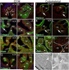
Figure 2. ECM and cytoskeletal proteins associated with human valve disease are disrupted in myxomatous ADAMTS5 deficient valves compared to WT
‘Intact versican’ GAGβ (green) in WT E15.5 (A) and the Adamts5−/− pulmonary valves (B). Collagen I localization at P1 in WT (C) and in Adamts5−/− valves (D, green; arrows (C, D) collagen I in the hinge region denoted by asterisk). Tropoelastin (green) localization at P1 in the ventricular aspect of the hinge in the WT (E) in contrast to the Adamts5−/− (F; open arrows (E, F) -elastin organization). Collagen III (green) at E15.5 is restricted to the spongiosa in the WT (G, line); in the Adamts5−/− valves collagen Ill is expanded in the cusp (H, line). Filamin A (green) in the hinge region of WT (I, K) and Adamts5−/− deficient valves at E17 and P8 respectively (J, L; arrows-differential staining of filamin A). αSMA expression (M, N, green) in P1 valves (arrowheads-differential αSMA expression). Electron micrographs of WT (O) and Adamts5−/− (P) valve hinge (P8; arrowheads- ECM intercell space). * (A-D, G-H)-hinge regions at the cusp-transient myocardial interface; Red-propidium iodide; blue- (A, B), αSMA; blue (G, H), α Sarcomeric actin. Scale bar in A = 100μm applies to B-H; I= 200μm applies to J-L; M = 50μm applies to N; O = 2 μm applies to P. n ≥ 4; each genotype.
Figure 3
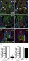
Figure 3. Smad2 is reduced in areas of versican accumulation in remodeling outflow tract cushions of ADAMTS5 deficient mice
pSmad2 (green) immunolocalization at E13.5 (A, B) and E12 (C, D) in WT (A, C) and Adamts5−/− (B, D) mesenchyme. ‘Intact versican’ GAGβ (green) (E, F) at E12 in sister sections of C and D respectively. Vertical bars denote areas with inversed correlation of versican and pSmad2. Arrows-subendocardial mesenchyme; asterisks-myocardium subjacent to the hinge; m-myocardium; D-distal outflow tract; P-proximal outflow tract. Red-propidium iodide; blue - (A-D) α-sarcomeric actin; blue - (E, F) αSMA. Quantification of pSmad2 nuclear staining E12-14 (G; n=4, WT, 4 different litters and Adamts5−/− hearts (n=4, Adamts5−/−, 4 different litters; * P < 1.5 × 10⁻⁷) normalized to WT. Quantification of pSmad2 nuclear staining E15.5-P8 (H; n=8, WT, 7 different litters and n=8, Adamts5−/−, 7 different litters; p<0.2950) normalized to WT. Scale Bar in A= 100μm and applies to B; C = 200μm and applies to D-F.
Figure 4
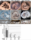
Figure 4. Adamts5−/−;Smad2+/− exhibit a high penetrance of bicuspid pulmonary and aortic valves
Dissected aortic valves (AV) at postnatal day 8 (P8; A-C) and 1 month (1mo; D-F). Arrowheads denote commissures (A-F) and highlight the anomalous and asymmetric commissure formation in Adamts5−/−;Smad2+/+ (B, E) and two versus three commissures in the Adamts5−/−; Smad2+/− (C, F). Opened AV (G-I) from adult hearts. Arrows (I) denote myxomatous bulges on the ventricular face of the bicuspid Adamts5−/−; Smad2+/− cusps. Graph (J) depicting phenotypic penetrance of bicuspid pulmonary and aortic valves of Adamts5−/−;Smad2+/− compared to intergenetic cross control genotypes. Dotted - BPV percentage; open bars - BAV percentage. Grey overlay -percentage of Adamts5−/−;Smad2+/− hearts that had both BPV and BAV. Scale bar in A = 350μm applies to B, C; D = 300 μm applies to E, F; G = 250μm applies to H and I.
Figure 5
Figure 5. Adamts5−/− mice with in vivo reduction of Smad2 display enlarged pulmonary and aortic valve cusps and hinge regions
Hematoxylin and eosin stained frontal sections of E17.5 pulmonary (PV) and aortic (AV) valves (A-F′). ′ -denotes section of the same heart, approximately 40 μm dorsally. Black arrowheads-hinge regions. Amira™ 3D reconstructions of E19.5 PV and AV cusps (G-L). L, red-left cusp of the PV; R, blue- right cusp of the PV; An, yellow- anterior cusp of the PV; purple-fusion of the R and L in the bicuspid PV; N, blue- non-coronary cusp of the AV; L, yellow-left coronary cusp of the AV; R, white- right coronary cusp of the AV; green- fusion of N and L of the bicuspid AV. Graph of average PV hinge (M) and cusp (N) width. Average width of AV hinge (O) and AV cusp (P). n=3, each genotype; p values as marked. *-denotes statistical significance. Scale bar in A = 150μm applies through F′; G = 200μm applies to H-L.
Figure 6
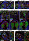
Figure 6. Endothelial nitrous oxide synthase (NOS3) is not significantly altered in the valvular endothelium of ADAMTS5 deficient mice
Expression of NOS3 was examined in histological sections of WT, Adamts5−/− and Adamts5−/−;Smad2+/− developing valves (A,B,C-E,F-G′ , green). NOS3 staining in valvular endothelium at E11.5 (A, B), E13.5 (C-E), and E14.5 (F, G), is depicted by arrowheads. Open arrowhead (B) shows altered pattern but not altered intensity of NOS3 in Adamts5−/−;Smad2+/−. Boxes (C-E; F, G) of the forming hinge region (mesenchymal-myocardial interface) shown in higher magnification in the inset (C-E) or in the adjacent panel (F′ and G′) and reveal mesenchymal NOS3 staining of the WT. NOS3 expression was noted in the arterial walls (open arrows C-E; F, G) Cleaved versican, i.e. DPEAAE staining (A′ and B′, green) present in Adamts5+/+;Smad2+/− and not detected in Adamts5−/−;Smad2+/− littermate. Intact versican (C′-E′, green) correlates with the hinge width and subjacent to the myocardium (C′-E′, blue). αSMA- alpha smooth muscle actin; αSarc-alpha sarcormeric actin. NOS3 immunohistochemistry involved E11.5-E15.5, n=4 each genotype. Scale bar in A = 100μm applies to A′, B, B′; C = 100μm applies to D, E and C,′ D′ and E′; Inset C = 20 μm and applies to all inset boxes; F = 200μm applies to G. Bar in F′ = 20μm applies to G′.
Figure 7
Figure 7. Schematic depicting versican cleavage in normal aortic and pulmonary valve development and its disruption in Adamts5−/− and Adamts5−/−;Smad2+/− mouse models with valve malformations
Whole hearts (A) depict the relative orientation of models in B and C; blue squares (A) show cross section (E11, E12), and oblique orientation (E13-E15) for models in B and C. Models depict WT (B) and ADAMTS5 deficient (C) development of outflow tract cushions. After septation (B, C, E13 E15) the predominant form of versican, either intact (royal blue) or DPEAAE (cleaved versican) (cyan) is shown in normal valve development (B), with a loss of DPEAAE (C). Malformations of valve development at specific stages are depicted (C). The bicuspid malformation, a predominant phenotype of the Adamts5−/−; Smad2+/− (C, E12) and delayed or insufficient excavation (C, E13, E15) resulting in enlarged hinge regions (C, E15) in Adamts5−/− and more severe in Adamts5−/−; Smad2+/− mice. Pink- neural crest cells (NCC) involved in outflow tract septation; Yellow- collagen fiber formation during ECM stratification (B, E15), reduced in Adamts5−/−; Smad2+/− and Adamts5−/− (C, E15) mice. White arrows (C, E12) show fusion of endocardial cells (EC) in bicuspid malformations of ADAMTS5 deficiency. Black arrowheads depict regions of normal excavation (B, E13, E15) or reduced excavation (C, E15); the developmental process of excavation creates the valvular sinus regions, which is the space between the valve cusps and the ascending arterial walls. Red-transient myocardium that expresses ADAMTS5; Orange- arterial wall tissue, defined as collagen-rich, α smooth muscle actin positive and α sarcormeric actin (myocardial marker) negative. LC- lateral outflow tract cushion; IC-intercalated cushion. Note: for simplicity we have left out NCC and endocardial derived cells however, a subset of these cells remain in the cusps at stages represented in these models.

Similar articles

Cited by

References

    1. Ward C. Clinical significance of the bicuspid aortic valve. Heart. 2000;83:81–5. - PMC - PubMed
    1. Calloway TJ, Martin LJ, Zhang X, Tandon A, Benson DW, Hinton RB. Risk factors for aortic valve disease in bicuspid aortic valve: a family-based study. Am J Med Genet A. 2011;155A:1015–20. - PubMed
    1. Supino PG, Borer JS, Yin A, Dillingham E, McClymont W. The epidemiology of valvular heart diseases: the problem is growing. Adv Cardiol. 2004;41:9–15. - PubMed
    1. Mordi I, Tzemos N. Bicuspid aortic valve disease: a comprehensive review. Cardiology research and practice. 2012;2012:196037. - PMC - PubMed
    1. Goda M, Budts W, Troost E, Meyns B. Bicuspid Pulmonary Valve With Atrial Septal Defect Leading to Pulmonary Aneurysm. Ann Thorac Surg. 2011;93:1706–8. - PubMed

MeSH terms