. 2013 Jul;33(7):1050-7.
doi: 10.1038/jcbfm.2013.46. Epub 2013 Mar 27.

Assessing white matter ischemic damage in dementia patients by measurement of myelin proteins

Affiliations

Assessing white matter ischemic damage in dementia patients by measurement of myelin proteins

Rachel Barker et al. J Cereb Blood Flow Metab. 2013 Jul.

Abstract

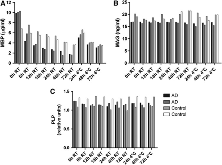
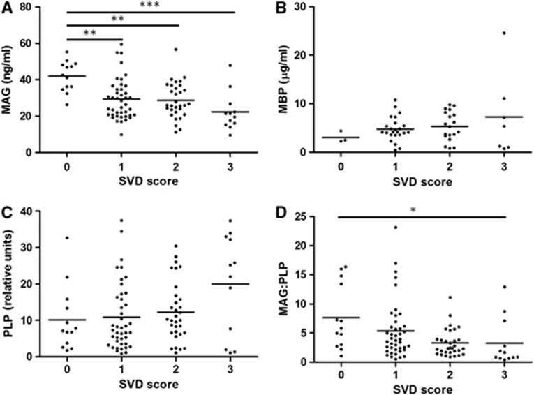
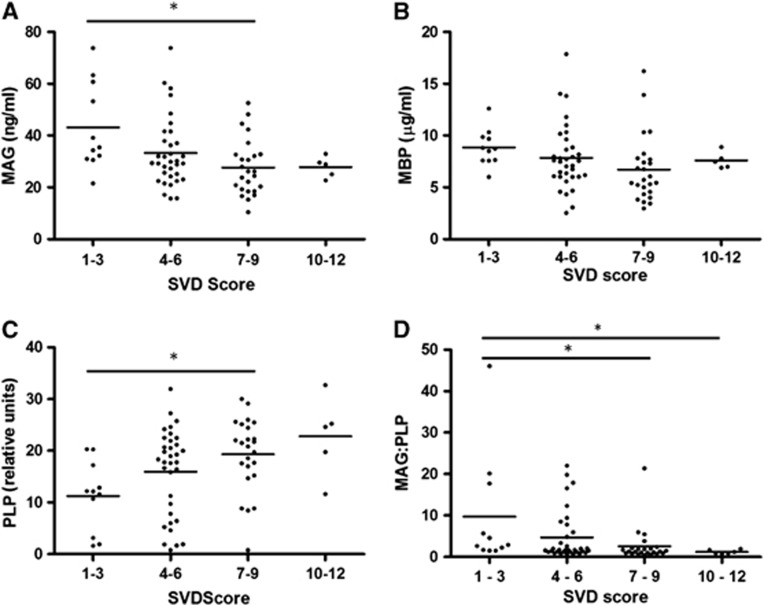
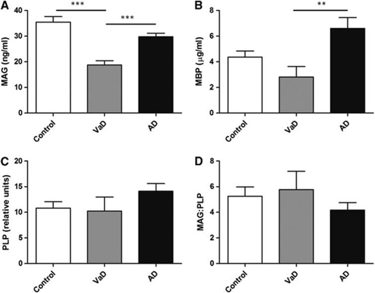
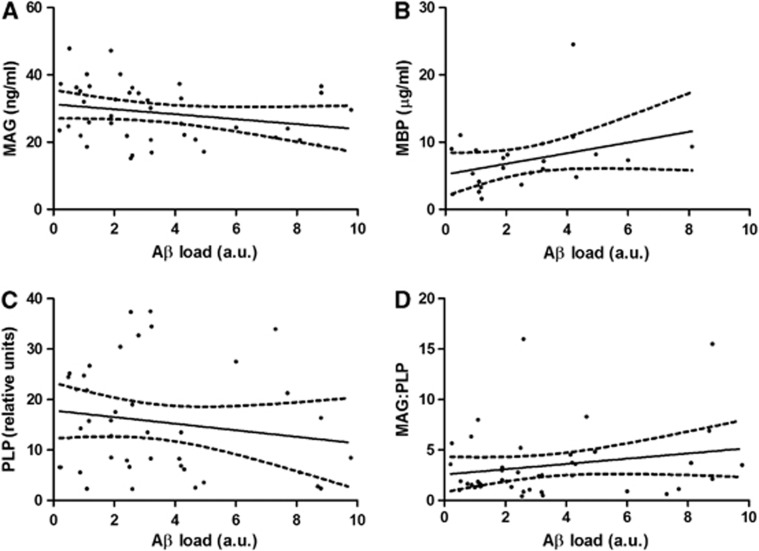
White matter ischemia is difficult to quantify histologically. Myelin-associated glycoprotein (MAG) is highly susceptible to ischemia, being expressed only adaxonally, far from the oligodendrocyte cell body. Myelin-basic protein (MBP) and proteolipid protein (PLP) are expressed throughout the myelin sheath. We compared MAG, MBP, and PLP levels in parietal white matter homogenates from 17 vascular dementia (VaD), 49 Alzheimer's disease (AD), and 33 control brains, after assessing the post-mortem stability of these proteins. Small vessel disease (SVD) and cerebral amyloid angiopathy (CAA) severity had been assessed in paraffin sections. The concentration of MAG remained stable post-mortem, declined with increasing SVD, and was significantly lower in VaD than controls. The concentration of MBP fell progressively post-mortem, limiting its diagnostic utility in this context. Proteolipid protein was stable post-mortem and increased significantly with SVD severity. The MAG/PLP ratio declined significantly with SVD and CAA severity. The MAG and PLP levels and MAG/PLP did not differ significantly between AD and control brains. We validated the utility of MAG and MAG/PLP measurements on analysis of 74 frontal white matter samples from an Oxford cohort in which SVD had previously been scored. MAG concentration and the MAG/PLP ratio are useful post-mortem measures of ante-mortem white matter ischemia.


Figures

Figure 1
Figure 1
Changes in myelin protein levels with post-mortem delay. Post-mortem delay was simulated by incubation of homogenates of adjacent samples of cortex at room temperature (RT) and 4°C for varying lengths of time. The protein levels of myelin-associated glycoprotein (MAG), myelin-basic protein (MBP), and proteolipid protein (PLP) were measured by enzyme-linked immunosorbent assay (ELISA). (A) The protein levels of MBP fell progressively with increasing incubation time, both at room temperature (r=−0.775, P=0.0408) and at 4°C (r=−0.951, P=0.0495). (B) The protein levels of MAG and (C) PLP did not alter significantly over 72 hours.
Figure 2
Figure 2
The relationship of (A) myelin-associated glycoprotein (MAG), (B) myelin-basic protein (MBP), (C) proteolipid protein (PLP), and the (D) MAG/PLP ratio to small vessel disease (SVD) in the deep parietal white matter in the SWDBB cohort. Both MAG and the MAG/PLP ratio correlated negatively with SVD severity (r=−0.384, P=0.0001 and r=−0.368, P=0.0002, respectively), whereas PLP correlated positively with SVD (r=0.305, P=0.0009). Myelin-associated glycoprotein was significantly reduced in cases with SVD severity of 1, 2 (**P<0.01), and 3 (***P<0.001) compared with those with no SVD. The MAG/PLP ratio was significantly reduced in cases with SVD severity of 3 compared with those with a score of 0 (*P<0.05). Myelin-basic protein did not vary significantly between the different SVD groups.
Figure 3
Figure 3
Double-immunofluorescent labeling of myelin-associated glycoprotein (MAG) and proteolipid protein (PLP) in the parietal white matter. Comparison between a case of high small vessel disease (SVD) severity (top panel) and one of low SVD severity (bottom panel) shows little difference in the labeling intensity of PLP but an obvious reduction in the labeling of MAG in severe SVD. Scale bar=100 μm.
Figure 4
Figure 4
The relationship of (A) myelin-associated glycoprotein (MAG), (B) myelin-basic protein (MBP), (C) proteolipid protein (PLP), and the (D) MAG/PLP ratio to small vessel disease (SVD) in the frontal white matter in the OPTIMA cohort. As in the SWDBB cohort, MAG and the MAG/PLP ratio declined significantly with increasing SVD score (r=−0.311, P=0.0066 and r=−0.349, P=0.0023, respectively) whereas PLP correlated positively with SVD severity (r=0.352, P=0.0021). Myelin-associated glycoprotein was significantly lower in cases with SVD severity of 7 to 9 than in those with severity of 1 to 3 (*P<0.05). The MAG/PLP ratio was significantly lower in cases with SVD severity of 7 to 9 and 10–12 than in those with severity of 1 to 3 (*P<0.05). Proteolipid protein was significantly higher in cases with SVD severity of 7 to 9 than those with severity of 1 to 3 (*P<0.05). Myelin-basic protein did not differ significantly between the SVD groups.
Figure 5
Figure 5
Levels of (A) myelin-associated glycoprotein (MAG), (B) myelin-basic protein (MBP), (C) proteolipid protein (PLP), and (D) the MAG/PLP ratio in parietal deep white matter from the SWDBB cohort in control, vascular dementia (VaD), and Alzheimer's disease (AD) brains. Myelin-associated glycoprotein concentration was significantly lower in VaD compared with controls and AD (***P<0.0001). Levels of MBP were reduced in VaD compared with AD (**P<0.01). PLP and the MAG/PLP ratio were not significantly different between the three groups.
Figure 6
Figure 6
Levels of (A) myelin-associated glycoprotein (MAG), (B) myelin-basic protein (MBP), (C) proteolipid protein (PLP), and (D) the MAG/:PLP ratio in parietal deep white matter from the SWDBB cohort, grouped according to cerebral amyloid angiopathy (CAA) severity. The levels of MAG, MBP, and PLP did not vary significantly with CAA. There was no significant difference in the ratio of MAG to PLP between the groups. However, MAG/PLP correlated negatively with the CAA severity score (r=−0.187, P=0.045).
Figure 7
Figure 7
The relationship of (A) myelin-associated glycoprotein (MAG), (B) myelin-basic protein (MBP), (C) proteolipid protein (PLP), and (D) the MAG/PLP ratio to parietal cortical amyloid-β (Aβ) plaque load in the Alzheimer's disease (AD) cohort. Levels of MAG were significantly negatively correlated with Aβ load (r=−0.318, P=0.0429) whereas MBP, PLP, and the MAG/PLP ratio were not significantly altered. The solid lines represent the best-fit linear regression lines, and the dashed lines the 95% confidence intervals. AU, arbitrary unit.

References

    1. Kalaria RN. The role of cerebral ischemia in Alzheimer's disease. Neurobiol Aging. 2000;21:321–330. - PubMed
    1. Meyer JS, Rauch GM, Rauch RA, Haque A, Crawford K. Cardiovascular and other risk factors for Alzheimer's disease and vascular dementia. Ann NY Acad Sci. 2000;903:411–423. - PubMed
    1. Whitmer RA, Sidney S, Selby J, Johnston SC, Yaffe K. Midlife cardiovascular risk factors and risk of dementia in late life. Neurology. 2005;64:277–281. - PubMed
    1. Luchsinger JA, Reitz C, Honig LS, Tang MX, Shea S, Mayeux R. Aggregation of vascular risk factors and risk of incident Alzheimer disease. Neurology. 2005;65:545–551. - PMC - PubMed
    1. Skoog I, Nilsson L, Persson G, Lernfelt B, Landahl S, Palmertz B, et al. 15-year longitudinal study of blood pressure and dementia. Lancet. 1996;347:1141–1145. - PubMed

Publication types

MeSH terms

Substances