. 2013 Jul;208(1):170-9.
doi: 10.1093/infdis/jit131. Epub 2013 Mar 26.

Expression of Pneumocystis jirovecii major surface glycoprotein in Saccharomyces cerevisiae

Affiliations

Expression of Pneumocystis jirovecii major surface glycoprotein in Saccharomyces cerevisiae

Geetha Kutty et al. J Infect Dis. 2013 Jul.

Abstract

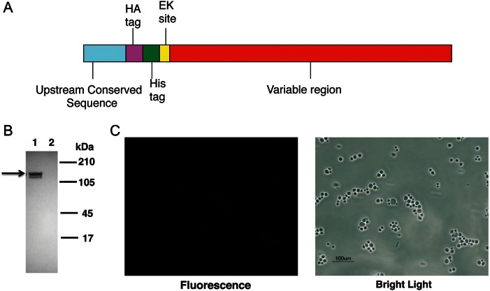
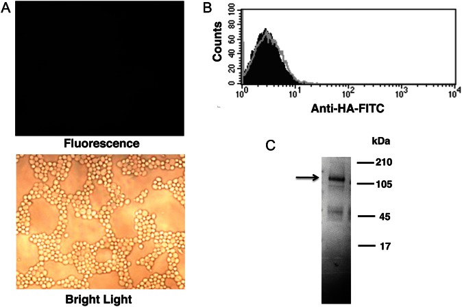
The major surface glycoprotein (Msg), which is the most abundant protein expressed on the cell surface of Pneumocystis organisms, plays an important role in the attachment of this organism to epithelial cells and macrophages. In the present study, we expressed Pneumocystis jirovecii Msg in Saccharomyces cerevisiae, a phylogenetically related organism. Full-length P. jirovecii Msg was expressed with a DNA construct that used codons optimized for expression in yeast. Unlike in Pneumocystis organisms, recombinant Msg localized to the plasma membrane of yeast rather than to the cell wall. Msg expression was targeted to the yeast cell wall by replacing its signal peptide, serine-threonine-rich region, and glycophosphatidylinositol anchor signal region with the signal peptide of cell wall protein α-agglutinin of S. cerevisiae, the serine-threonine-rich region of epithelial adhesin (Epa1) of Candida glabrata, and the carboxyl region of the cell wall protein (Cwp2) of S. cerevisiae, respectively. Immunofluorescence analysis and treatment with β-1,3 glucanase demonstrated that the expressed Msg fusion protein localized to the yeast cell wall. Surface expression of Msg protein resulted in increased adherence of yeast to A549 alveolar epithelial cells. Heterologous expression of Msg in yeast will facilitate studies of the biologic properties of Pneumocystis Msg.

Keywords: GPI anchored protein; Pneumocystis jirovecii; antigenic variation; major surface glycoprotein; upstream conserved sequence.


Figures

Figure 1.
Figure 1.
Recombinant Msg protein expressed in Saccharomyces cerevisiae does not localize to the cell wall. A, Schematic diagram of the Pneumocystis jirovecii msg construct that was inserted in pESC-URA vector. The hemagglutinin (HA) tag, histidine (His) tag, and enterokinase site were added between the upstream conserved sequence and variable region of msg. B, Western blot analysis of expressed protein, using peroxidase-conjugated anti-HA antibody. Protein extracts prepared from cells transformed with msg construct (lane 1) showed immunoreactivity to 2 approximately 130-kDa bands (arrow), while extracts from cells transformed with vector alone (lane 2) showed no immunoreactivity. C, Immunofluorescence microscopy analysis of yeast cells expressing Msg using FITC-conjugated anti-HA antibody. The cells expressing Msg showed no fluorescence when viewed under UV light, indicating that Msg is not expressed on the yeast cell wall. Yeast cells can be seen under bright light.
Figure 2.
Figure 2.
Expressed Msg is localized to the plasma membrane of Saccharomyces cerevisiae and is glycosylated. Yeast cells transformed with Msg or vector alone were treated with β-1,3 glucanase, and the supernatant containing the digested cell wall and the pellet containing spheroplasts were separated by centrifugation. Spheroplasts were further fractionated to yield cell membranes and the cytosol. A, Western blot analysis of the cell fractions, using anti-hemagglutinin (HA) antibody. Spheroplasts (lane 2) and the cell membrane (lane 3) fraction showed immunoreactivity to 2 approximately 130-kDa bands (arrow). No immunoreactivity was seen with digested cell wall (lane 1) or the cytosol preparation (lane 4). B, Immunofluorescence microscopic analysis of spheroplasts using fluorescein isothiocyanate–conjugated anti-HA antibody. Spheroplasts prepared from yeast cells expressing Msg showed fluorescence, while cells transformed with vector alone showed no fluorescence. C, Western blot analysis of Msg before and after deglycosylation. Proteins extracted from the cell membrane fraction, which contains Msg, were treated with pNGase F prior to Western blot analysis using anti-HA antibody. Untreated Msg migrates as 2 close bands (lane 1); following deglycosylation, there is a single band (lane 2) that is slightly smaller than the higher molecular weight band seen in lane 1.
Figure 3.
Figure 3.
Mutation of amino acids at the putative ω-site region of Pneumocystis jirovecii Msg does not alter the localization of expressed Msg. A, Alignment of C-terminal amino acid residues of P. jirovecii Msg with that of yjr151c, a glycophosphatidylinositol-dependent cell wall protein of Saccharomyces cerevisiae. The protein sequences for the 2 mutated constructs are shown below the alignment. The hydrophobic amino acid residues and the amino acid at the potential ω-site are shown in bold. The mutated amino acids are shown in bold and underlined. B, Western blot analysis using anti-hemagglutinin (HA) antibody. Cells transformed with the original construct (lane 1), mutation 1 (lane 2), and mutation 2 (lane 3) all expressed recombinant Msg in yeast, as shown by immunoreactivity to an approximately 130-kDa band (arrow) in Western blot analysis using anti-HA antibody, but showed no surface reactivity by immunofluorescent staining (data not shown).
Figure 4.
Figure 4.
Targeting Msg to the yeast cell wall using pBC542 vector. A fragment of the msg gene was cloned into pBC542 vector, which contains sequences encoding a serine-threonine–rich region of Epa1 of Candida glabrata followed by sequences encoding the carboxyl region of Cwp2 of Saccharomyces cerevisiae. A, The schematic diagram of 3 DNA constructs used for expression in pBC542. The Msg fragment (1–2771 bp of msg 32) excludes a serine-threonine–rich region and the glycophosphatidylinositol anchor signal of Msg. Construct 1 includes the entire upstream conserved sequence (UCS) of Msg, as well as most of the variable region. In construct 2, the Msg signal peptide in construct 1 was replaced by a yeast signal peptide. In construct 3, the UCS sequence through the predicted cleavage site was removed from construct 2, while the yeast signal peptide was retained. B, Immunofluorescence microscopic analysis of yeast transformants. Cells transformed with constructs 2 and 3 showed immunofluorescence when stained with fluorescein isothiocyanate (FITC)–conjugated anti-hemagglutinin (HA) antibody, suggesting cell wall localization of expressed Msg fusion protein. No fluorescence was detected when cells were transformed with construct 1. C, Flow cytometric analysis using FITC-conjugated anti-HA antibody. FITC-conjugated nonspecific antibody (mouse immunoglobulin G) was used as negative control. The histogram shows the cell counts on the y-axis and fluorescence intensity on the x-axis. The solid histogram shows nonspecific staining with the negative control, and the open histogram shows specific staining with the anti-HA antibody. The staining of cells transformed with construct 1 was similar to that of the negative control, while cells transformed with construct 2 and construct 3 showed immunofluorescence staining of approximately 70% of the cells. D, Western blot analysis of protein extracts prepared from cells following disruption with glass beads. Cells expressing construct 1 showed immunoreactivity to an approximately 150-kDa band (expected size is shown by the arrow) when probed with anti-HA antibody (lane 1), while cells expressing construct 2 (lane 2) and construct 3 (lane 3) showed a high molecular weight smear (bracket), indicating that the protein is still attached to the cell wall. E, Immunofluorescence and Western blot analysis of protein extracts prepared from cells expressing construct 3 following treatment with β-1,3 glucanase. Immunofluorescence reactivity in untreated cells is lost following β-1,3 glucanase treatment. Glucanase treatment solubilizes recombinant Msg, which now migrates as an approximately 150-kDa band (arrow), rather than as the high molecular weight smear that was seen following glass bead disruption.
Figure 5.
Figure 5.
Replacing the Epa1 serine–threonine–rich region with a serine-threonine–rich region of Msg disrupts cell wall localization of Msg fusion protein. The Epa1 sequence of construct 2 (Figure 4) was replaced by a sequence encoding 4 repeats (59 amino acids each) of the Msg serine-threonine–rich region. Protein expression in transformed yeast cells was analyzed by immunofluorescence and Western blot. A, Immunofluorescence microscopic analysis using fluorescein isothiocyanate (FITC)–conjugated anti-hemagglutinin (HA) antibody showed no surface fluorescence, while multiple organisms are seen by bright light. B, Flow cytometric analysis of yeast cells stained with FITC-conjugated anti-HA antibody. The histogram shows the cell counts on the y-axis and fluorescence intensity on the x-axis. Solid histogram shows nonspecific staining with the negative control (mouse immunoglobulin G), and the open histogram shows specific staining with the anti-HA antibody. Consistent with the microscopic results, no specific staining is seen. C, By Western blot analysis, an approximately 150-kDa band is seen when probed with anti-HA antibody, demonstrating that the fusion protein is expressed.
Figure 6.
Figure 6.
Improved cell surface expression of Msg fusion protein under the control of gal1 promoter. Construct 3 (Figure 4A) was cloned into pESC-URA vector downstream of the gal1 promoter. After 4 hours of galactose induction, the yeast transformants were analyzed for the cell surface expression of Msg by immunofluorescence, using fluorescein isothiocyanate–conjugated anti-hemagglutinin (HA) antibody. A, Immunofluorescence microscopic analysis. Cells transformed with the msg construct showed immunofluorescence, whereas cells transformed with vector alone showed no fluorescence. B, Flow cytometric analysis. The histogram shows the cell counts on the y-axis and fluorescence intensity on the x-axis. Solid histogram shows nonspecific staining with the negative control (mouse immunoglobulin G), and the open histogram shows specific staining with the anti-HA antibody. Approximately 84% of the cells showed surface expression of the Msg fusion protein; vector alone showed no staining.
Figure 7.
Figure 7.
Yeast cells expressing Msg on the cell surface adhere to A549 epithelial cells. Cells transformed with msg construct 3 expressed in pESC-URA or vector alone were stained with carboxyfluorescein diacetate, succinimidyl ester (CFSE) and then tested for their ability to adhere to cultured A549 epithelial cells. A, Fluorescence microscopic analysis demonstrates that most of the yeast cells visible under bright light are stained with CFSE. B, Yeast cells expressing Msg show increased adherence to epithelial cells. After 1 hour of incubation with CSFE-labeled yeast, followed by washing, epithelial cells and adherent yeast were scraped, and fluorescence was quantitated with a fluorometer. Results are the mean of 2 experiments. The error bar represents the SD. Although the results are not significant, in both experiments yeast cells expressing Msg showed an approximately 9-fold higher fluorescence as compared to those transformed with vector alone.

Similar articles

Cited by

References

    1. Kovacs JA, Masur H. Evolving health effects of Pneumocystis: one hundred years of progress in diagnosis and treatment. JAMA. 2009;301:2578–85. - PubMed
    1. Thomas CF, Jr, Limper AH. Pneumocystis pneumonia. N Engl J Med. 2004;350:2487–98. - PubMed
    1. Stringer JR, Stringer SL, Zhang J, Baughman R, Smulian AG, Cushion MT. Molecular genetic distinction of Pneumocystis carinii from rats and humans. J Eukaryot Microbiol. 1993;40:733–41. - PubMed
    1. Sinclair K, Wakefield AE, Banerji S, Hopkin JM. Pneumocystis carinii organisms derived from rat and human hosts are genetically distinct. Mol Biochem Parasitol. 1991;45:183–4. - PubMed
    1. Mazars E, Guyot K, Durand I, et al. Isoenzyme diversity in Pneumocystis carinii from rats, mice, and rabbits. J Infect Dis. 1997;175:655–60. - PubMed

Publication types

MeSH terms

Substances