. 2013 May 10;112(10):1334-44.
doi: 10.1161/CIRCRESAHA.113.301055. Epub 2013 Mar 26.

Focal energy deprivation underlies arrhythmia susceptibility in mice with calcium-sensitized myofilaments

Affiliations

Focal energy deprivation underlies arrhythmia susceptibility in mice with calcium-sensitized myofilaments

Sabine Huke et al. Circ Res. .

Abstract

Rationale: The Ca(2+) sensitivity of the myofilaments is increased in hypertrophic cardiomyopathy and other heart diseases and may contribute to a higher risk for sudden cardiac death. Ca(2+) sensitization increases susceptibility to reentrant ventricular tachycardia in animal models, but the underlying mechanism is unknown.

Objective: To investigate how myofilament Ca(2+) sensitization creates reentrant arrhythmia susceptibility.

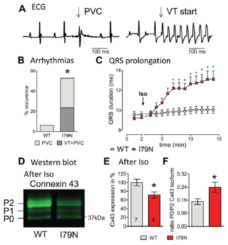
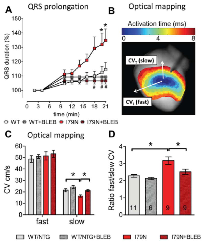
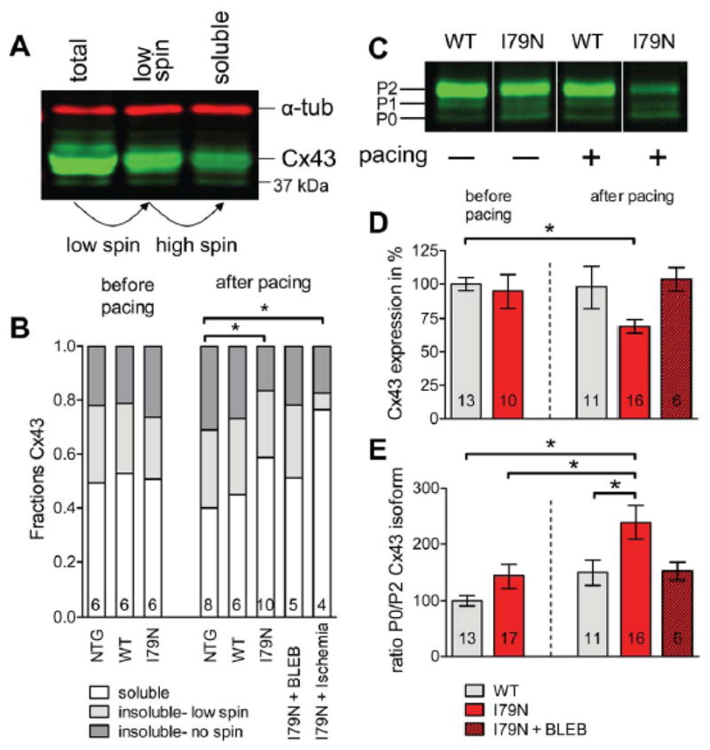
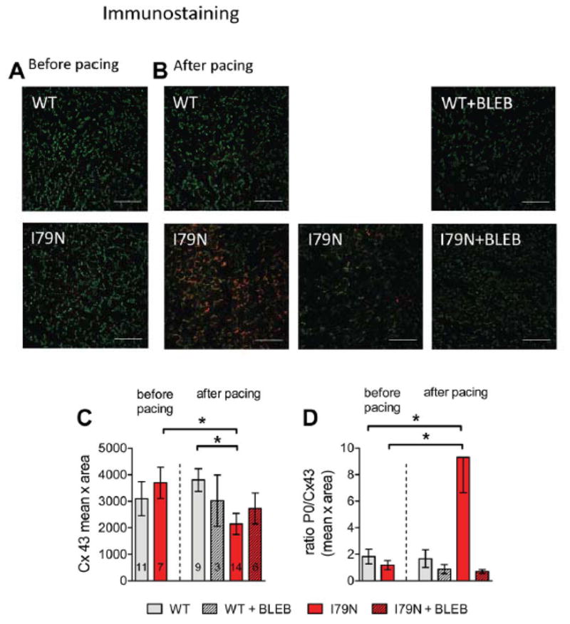
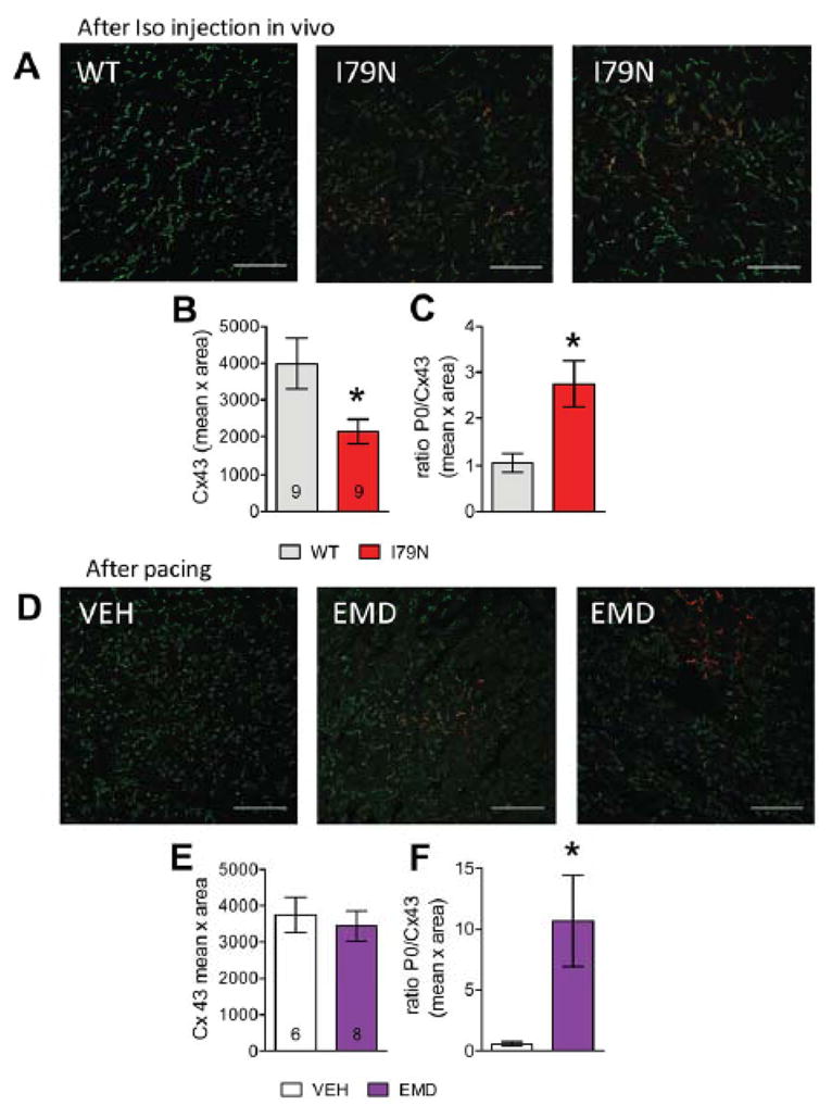
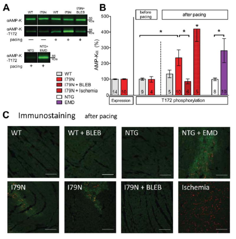
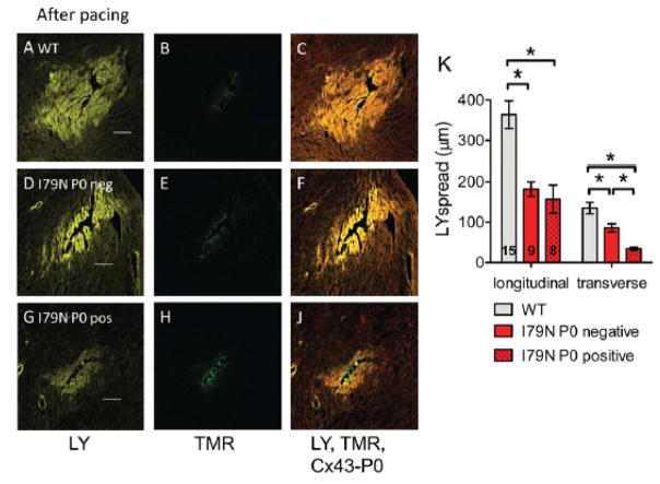
Methods and results: Using hypertrophic cardiomyopathy mouse models (troponinT-I79N) and a Ca(2+) sensitizing drug (EMD57033), here we identify focal energy deprivation as a direct consequence of myofilament Ca(2+) sensitization. To detect ATP depletion and thus energy deprivation, we measured accumulation of dephosphorylated Connexin 43 (Cx43) isoform P0 and AMP kinase activation by Western blotting and immunostaining. No differences were detected between groups at baseline, but regional accumulation of Connexin 43 isoform P0 occurred within minutes in all Ca(2+)-sensitized hearts, in vivo after isoproterenol challenge and in isolated hearts after rapid pacing. Lucifer yellow dye spread demonstrated reduced gap junctional coupling in areas with Connexin 43 isoform P0 accumulation. Optical mapping revealed that selectively the transverse conduction velocity was slowed and anisotropy increased. Myofilament Ca(2+) desensitization with blebbistatin prevented focal energy deprivation, transverse conduction velocity slowing, and the reentrant ventricular arrhythmias.

Conclusions: Myofilament Ca(2+) sensitization rapidly leads to focal energy deprivation and reduced intercellular coupling during conditions that raise arrhythmia susceptibility. This is a novel proarrhythmic mechanism that can increase arrhythmia susceptibility in structurally normal hearts within minutes and may, therefore, contribute to sudden cardiac death in diseases with increased myofilament Ca(2+) sensitivity.

Keywords: Connexin 43; conduction velocity dispersion; familial hypertrophic cardiomyopathy; myofilament Ca2+ sensitivity; sudden cardiac death; ventricular arrhythmias.


Figures

Fig. 1
Fig. 1. Isoproterenol (Iso) injection in vivo induces ventricular arrhythmias, slowed ventricular depolarization and is associated with reduced expression of the gap junction protein Cx43 in TnT-I79N mice
(A) Representative examples for a premature ventricular contraction (PVC) and ventricular tachycardia (VT). Summary data are presented in (B), n=16 each. (C) QRS duration was prolonged within a few minutes after Iso injection in TnT-I79N, but not TnT-WT mice. (D) Typical Western blot pattern for anti-Cx43 (isoforms P2, P1, P0 indicated) after Iso treatment, (E) Total expression of Cx43 and (F) P0/P2 isoform ratio analyzed by Western blot. N numbers are indicated in columns and are the same for (E) and (F). * p≤0.05 vs WT
Fig. 2
Fig. 2. Isolated TnT-I79N hearts exhibit QRS prolongation and slower transverse conduction velocity (CVT)
(A) QRS duration determined from volume conducted ECG recordings during brief pacing interruptions (pacing protocol as shown in Supplemental Fig. I); n = 6–19. QRS prolongation was observed in TnT-I79N, but not in TnT-WT and was prevented by blebbistatin (BLEB 3 μM, n=3–18). (C) Longitudinal (fast, CVL) and CVT (slow) conduction velocity and (D) anisotropy ratio in control (WT/NTG) and I79N determined as illustrated in (B). No difference was observed between TnT-WT and NTG mice and data were pooled. N numbers are indicated in columns and are the same for (C) and (D). * p≤0.05; (A) * p≤0.05 vs WT, # p≤0.05 vs I79N
Fig. 3
Fig. 3. Rapid pacing induces changes in connexin43 (Cx43) protein expression, solubility and isoform distribution in TnT-I79N isolated hearts (pacing protocol see Supplemental Fig. IA)
(B) Fractions of soluble (non-junctional) and insoluble (junctional) Cx43 in NTG, WT and I79N. N numbers are indicated in columns. A representative Western blot is shown (A, α-tubulin (α-tub)). The same sample volume was loaded before centrifugation (total), after 500×g centrifugation (low spin) and after 14000×g centrifugation (soluble). (C) A representative Western blot (all samples from same blot, but intermediate lanes were deleted) of WT and I79N samples before and after pacing. The different Cx43 isoforms are indicated (P2, P1, P0). (D) Summary data for Cx43 expression and (E) Summary data for the ratio of P0/P2 isoform for WT and I79N before and after pacing and I79N after pacing in the presence of BLEB (3 μM). * p≤0.05
Fig. 4
Fig. 4
Regional accumulation of dephosphorylated Cx43 isoform P0 in TnT-I79N after pacing. This is not observed in TnT-WT, in TnT-I79N before pacing and is prevented by BLEB. Anti-Cx43 (green) and anti-Cx43-P0 (red) staining of I79N heart sections before (A) and after pacing (B, two examples are shown for I79N after pacing). The scale bar length is 100 μm. (C) Summary data for integrated density (mean intensity x area (pixel)) of Cx43 (N numbers are indicated in columns and are the same for both graphs). See Supplemental Fig. III for examples of anti-Cx43 staining (green) only for all groups. (D) Summary data for the average ratio of Cx43/Cx43-P0 in all groups (C) * p≤0.05
Fig. 5
Fig. 5. Regional accumulation of Cx43-P0 observed in I79N after Iso injection in vivo and after treatment of isolated control hearts with the Ca2+ sensitizer EMD57033 (EMD, 3 μM)
(A) Anti-Cx43 (green) and anti-Cx43-P0 (red) staining of TnT-WT and TnT-79N ventricular tissue after Iso challenge. (B) Summary data for integrated density (mean intensity x area (pixel)) of Cx43 and (C) ratio of Cx43-P0/Cx43. (D) Examples of immunostained tissue from isolated NTG hearts treated with VEH or EMD after rapid pacing. (E+F) Summary data for EMD experiments (for pacing protocol see Supplemental Fig. IA). The scale bar length is100 μm in all images. * p≤0.05 vs WT
Fig. 6
Fig. 6. Activated AMP-kinase (pAMPK) in areas with dephosphorylated Cx43-P0 accumulation
(A+B) Western blot analysis of αAMP-K expression and phosphorylation at threonine 172 (=activation). Representative western blots (A, white line indicates where intermediate lanes were deleted) and summary data (B)). 21 min of global ischemia was included as a positive control. (C) Representative examples showing activated pAMPK –T172 (“green”) in the same regions where Cx43-P0 accumulation (“red”) is observed (TnT-I79N, bottom first and second and NTG+EMD, top forth). Cx43-P0 is more uniformly observed after global ischemia (bottom forth). The scale bar indicates 100 μm. * p≤0.05 as indicated.
Fig. 7
Fig. 7. Reduced gap junctional coupling assessed by modified scrape loading
(A,D,G) Representative confocal images of Lucifer yellow (LY, yellow) spread after needle puncture in TnT hearts after pacing. (B,E,H) Corresponding images of non-gap junction permeable Tetramethylrhodamine dextran (TMR, green) that was simultaneously injected. (C, F, J) Composite image of LY, TMR and Cx43-P0 (red). The scale bar length is 100 μm. (K) Summary data of LY spread analysis from 15 sites in TnT-WT (3 hearts) and 17 sites in TnT-I79N hearts (4 hearts). For TnT-I79N only sites were analyzed that were clearly classified as Cx43-P0 negative (I79N P0 neg, n=9) or showed clear Cx43-P0 accumulation along at least two sides (I79N P0 pos, n=8). Dye spread was analyzed along the fiber (longitudinal) or across the fiber (transverse). * p≤0.05 vs. as indicated

References

    1. Maron BJ, Gardin JM, Flack JM, Gidding SS, Kurosaki TT, Bild DE. Prevalence of hypertrophic cardiomyopathy in a general population of young adults. Echocardiographic analysis of 4111 subjects in the CARDIA Study. Coronary Artery Risk Development in (Young) Adults. Circulation. 1995;92:785–789. - PubMed
    1. Keren A, Syrris P, McKenna WJ. Hypertrophic cardiomyopathy: the genetic determinants of clinical disease expression. Nat Clin Pract Cardiovasc Med. 2008;5:158–168. - PubMed
    1. Maron BJ, Olivotto I, Spirito P, Casey SA, Bellone P, Gohman TE, Graham KJ, Burton DA, Cecchi F. Epidemiology of hypertrophic cardiomyopathy-related death: revisited in a large non-referral-based patient population. Circulation. 2000;102:858–864. - PubMed
    1. Shirani J, Pick R, Roberts WC, Maron BJ. Morphology and significance of the left ventricular collagen network in young patients with hypertrophic cardiomyopathy and sudden cardiac death. J Am Coll Cardiol. 2000;35:36–44. - PubMed
    1. Elliott P, Spirito P. Prevention of hypertrophic cardiomyopathy-related deaths: theory and practice. Heart. 2008;94:1269–1275. - PubMed

Publication types

MeSH terms