

Angiotensin II Receptor Antagonism Reduces Transforming Growth Factor Beta and Smad Signaling in Thoracic Aortic Aneurysm
- PMID: 23532685
- PMCID: PMC3603187
Angiotensin II Receptor Antagonism Reduces Transforming Growth Factor Beta and Smad Signaling in Thoracic Aortic Aneurysm
Abstract
Background: The expression of transforming growth factor beta (TGF-β) and Smad3 regulates extracellular matrix homeostasis and inflammation in aortic aneurysms. The expression of Smad3 depends on signaling by angiotensin II (AngII) receptor pathways through TGF-β receptor-dependent and -independent pathways.
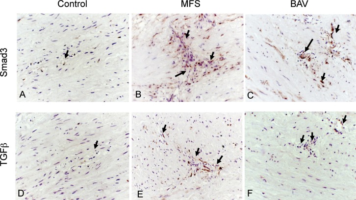
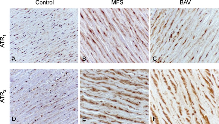
Methods: To determine the expression of AngII type 1 (AT1R) and type 2 receptors (AT2R), TGF-β, and Smad3 in thoracic aortic aneurysms, we performed immunohistochemistry testing on tissue and cultured cells derived from subjects with Marfan syndrome (MFS) and bicuspid aortic valve (BAV) malformation and from normal aortas of subjects who were organ donors.
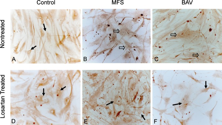
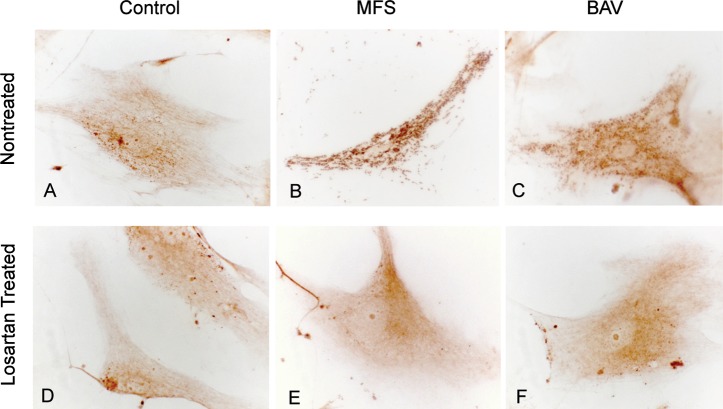
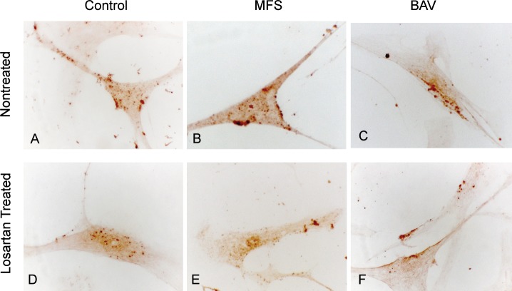
Results: MFS and BAV aneurysm tissue showed enhanced accumulation of TGF-β and Smad3 in vascular smooth muscle cells (VSMCs) and in inflammatory cells in the subintimal layer and tunica media. The normal aortic wall exhibited minimal TGF-β and Smad3 staining. Cultured VSMCs from MFS and BAV samples showed nuclear Smad3 and strong cytoplasmic TGF-β expression in the cytoplasmic vesicles. In control cells, Smad3 was located mainly in the cytoplasm, and weak cytoplasmic TGF-β was distributed with a pattern similar to that of the aneurysm-derived cells. Compared to normal aorta cells, AT1R and AT2R expression was increased in both aneurysm types. Treatment of cultured VSMCs with the AT1R antagonist losartan caused both reduced TGF-β vesicle localization and nuclear expression of Smad3.
Conclusions: Increased TGF-β and Smad3 expression in aneurysm tissue and cultured VSMCs is consistent with aberrant TGF-β expression and the activation of Smad3 signaling. Losartan-mediated reduction in TGF-β expression and the cytoplasmic localization of Smad3 support a role for AT1R antagonism in the inhibition of aneurysm progression.
Keywords: Aneurysm; Smad3 protein; aorta; immunohistochemistry; transforming growth factor beta; vascular smooth muscle.
Conflict of interest statement
Figures
References
-
- Ince H, Nienaber CA. Etiology, pathogenesis and management of thoracic aortic aneurysm. Nat Clin Pract Cardiovasc Med. 2007 Aug;4(8):418–427. - PubMed
-
- Kaartinen V, Warburton D. Fibrillin controls TGF-beta activation. Nat Genet. 2003 Mar;33(3):331–332. - PubMed
-
- Bobik A. Transforming growth factor-betas and vascular disorders. Arterioscler Thromb Vasc Biol. 2006 Aug;26(8):1712–1720. Epub 2006 May 4. - PubMed
-
- Heldin CH, Miyazono K, ten Dijke P. TGF-beta signalling from cell membrane to nucleus through SMAD proteins. Nature. 1997 Dec 4;390(6659):465–471. - PubMed
LinkOut - more resources
Full Text Sources