. 2013 May 10;288(19):13305-16.
doi: 10.1074/jbc.M113.457937. Epub 2013 Mar 26.

Calcium-dependent phospholipid scramblase activity of TMEM16 protein family members

Affiliations

Calcium-dependent phospholipid scramblase activity of TMEM16 protein family members

Jun Suzuki et al. J Biol Chem. .

Abstract

Background: TMEM16A and 16B work as Cl(-) channel, whereas 16F works as phospholipid scramblase. The function of other TMEM16 members is unknown.

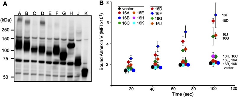
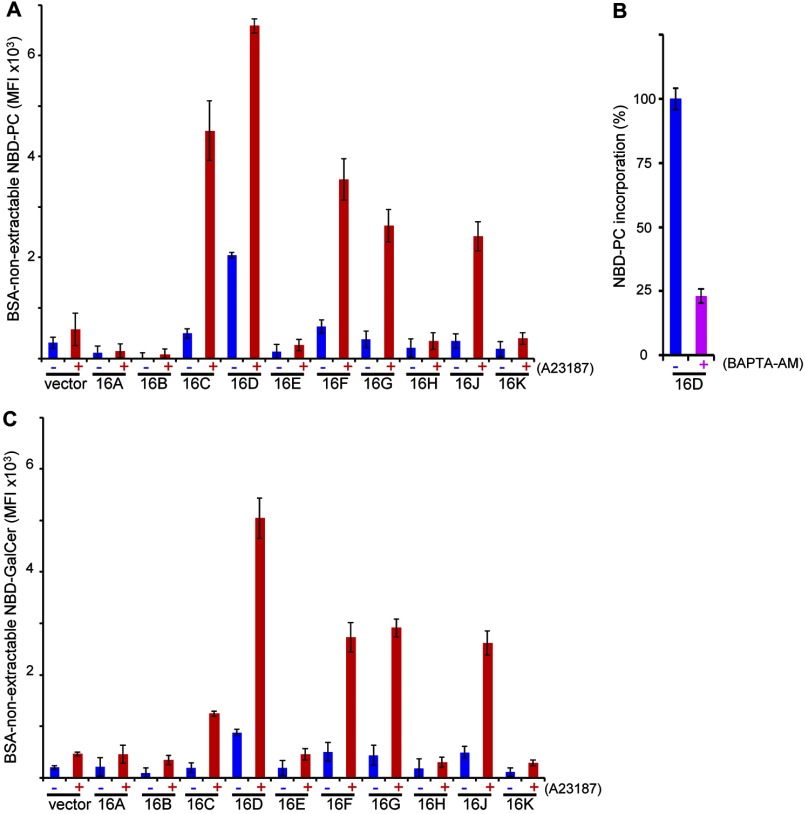
Results: Using TMEM16F(-/-) cells, TMEM16C, 16D, 16F, 16G, and 16J were shown to be lipid scramblases.

Conclusion: Some TMEM16 members are divided into two Cl(-) channels and five lipid scramblases.

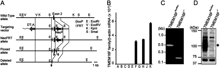
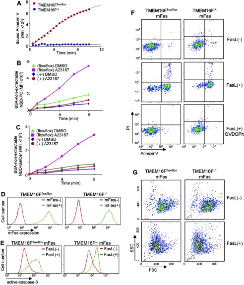
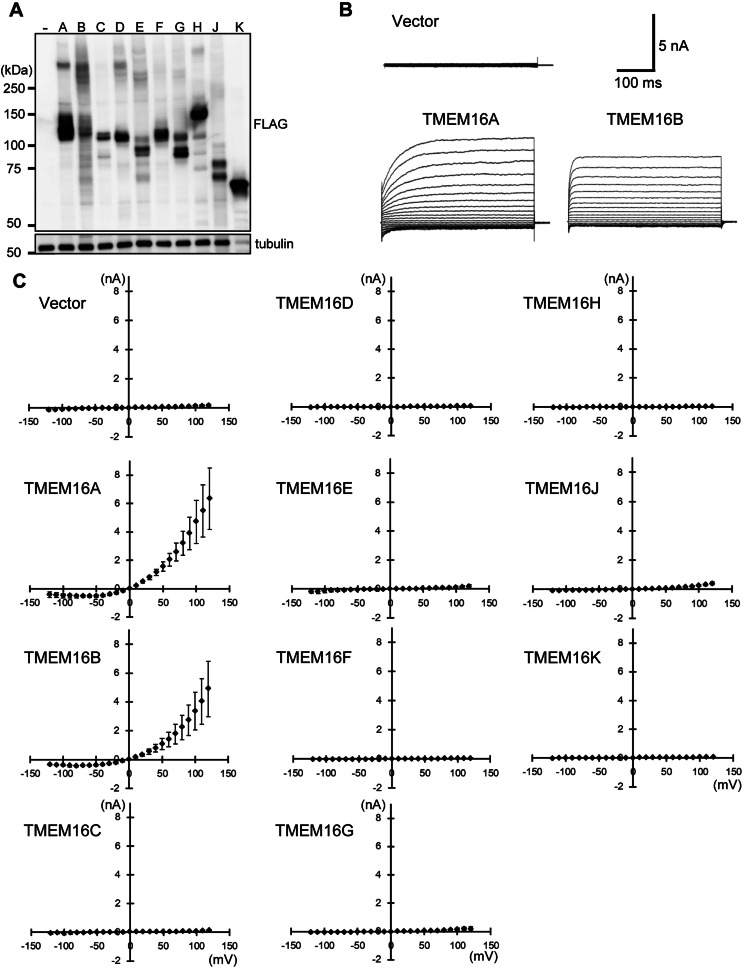
Significance: Learning the biochemical function ofTMEM16family members is essential to understand their physiological role. Asymmetrical distribution of phospholipids between the inner and outer plasma membrane leaflets is disrupted in various biological processes. We recently identified TMEM16F, an eight-transmembrane protein, as a Ca(2+)-dependent phospholipid scramblase that exposes phosphatidylserine (PS) to the cell surface. In this study, we established a mouse lymphocyte cell line with a floxed allele in the TMEM16F gene. When TMEM16F was deleted, these cells failed to expose PS in response to Ca(2+) ionophore, but PS exposure was elicited by Fas ligand treatment. We expressed other TMEM16 proteins in the TMEM16F(-/-) cells and found that not only TMEM16F, but also 16C, 16D, 16G, and 16J work as lipid scramblases with different preference to lipid substrates. On the other hand, a patch clamp analysis in 293T cells indicated that TMEM16A and 16B, but not other family members, acted as Ca(2+)-dependent Cl(-) channels. These results indicated that among 10 TMEM16 family members, 7 members could be divided into two subfamilies, Ca(2+)-dependent Cl(-) channels (16A and 16B) and Ca(2+)-dependent lipid scramblases (16C, 16D, 16F, 16G, and 16J).

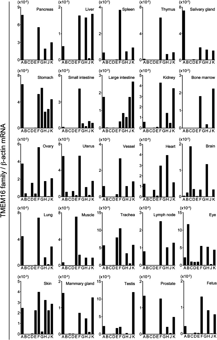
Keywords: Calcium; Cl- Channel; Lipid Transport; Phosphatidylcholine; Phosphatidylserine; Phospholipid Scramblase; Plasma Membrane; TMEM16; Tissue-specific Expression.


Figures

FIGURE 1.
FIGURE 1.
Establishment of TMEM16F−/− IFET cell line. A, schematic representation of wild-type and mutant TMEM16F alleles together with the targeting vector. Recognition sites for EcoRI (E), EcoRV (V), KpnI (K), and SmaI (S) in the flanking region of exon 2 (filled boxes) are indicated. In the target vector, a 1.0-kb DNA fragment carrying exon 2 and its flanking region were replaced by a 2.7-kb fragment carrying two loxP sequences (filled arrowheads) and PGK-neo (NeoR) flanked by FRT sequences (gray arrowheads). Diphtheria toxin A fragment (DT-A) driven by the tk promoter was inserted at the 5′ site of the vector. In NeoFRT allele, the TMEM16F chromosomal gene was replaced by the targeting vector. In Floxed allele, the FRT-flanked NeoR gene was removed by FLPe recombinase. In deleted allele, the loxP-flanked exon 2 of TMEM16F gene was deleted by Cre recombinase. Primers used in C are indicated by arrows. Scale bar, 1.0 kb. B, real-time PCR analysis for mRNA of TMEM16F family members in IFETs. An IFET cell line was established from TMEM16Fflox/flox fetal thymocytes. TMEM16A–16H, 16J, and 16K mRNA in TMEM16Fflox/flox IFETs was quantified by real-time PCR and expressed relative to β-actin mRNA. The experiment was carried out for three times, and the average value was plotted ± S.D. (error bars). C, deletion of TMEM16F exon 2 in the IFET cell line. TMEM16Fflox/flox IFETs were infected by Cre-bearing adenovirus to establish TMEM16F−/− IFET cells. Chromosomal DNA from TMEM16Fflox/flox and TMEM16F−/− IFETs was analyzed by PCR with the primers indicated in A. D, Western blots for TMEM16F in TMEM16Fflox/flox and TMEM16F−/− IFETs. Cell lysates (10 μg of proteins) were separated by 7.5% SDS-PAGE and blotted with rabbit anti-TMEM16F serum (upper panel) or anti-α-tubulin antibody (lower panel). Molecular mass standards (Precision Plus Standard; Bio-Rad) are shown in kDa at left.
FIGURE 2.
FIGURE 2.
An indispensable role of TMEM16F for Ca2+-induced but not apoptotic PS exposure. A, Ca2+ ionophore-induced PS exposure. TMEM16Fflox/flox and TMEM16F−/− IFETs were treated at 20 °C with 3.0 μm A23187 in the presence of Cy5-labeled annexin V. Annexin V binding to the cells was monitored by flow cytometry for 10 min and expressed in mean fluorescence intensity (MFI). B and C, Ca2+ ionophore-induced lipid internalization. TMEM16Fflox/flox and TMEM16F−/− IFETs were treated at 15 °C with 250 nm A23187 in the presence of 100 nm NBD-PC (B) or 250 nm NBD-GalCer (C). Using aliquots of the reaction mixture, the BSA-nonextractable level of NBD-PC or NBD-GalCer in the Sytox Blue-negative population was determined at the indicated time by FACSAria and expressed in mean fluorescence intensity. D, transformation of IFETs with mouse Fas. TMEM16Fflox/flox and TMEM16F−/− IFETs were infected with a retrovirus carrying mouse Fas and were stained with a PE-labeled hamster mAb against mouse Fas (green). The staining profile of parental cells is also shown (red). E–G, FasL-induced apoptosis. Fas-expressing TMEM16Fflox/flox and TMEM16F−/− IFETs were treated at 37 °C for 2 h with 60 units/ml FasL in the absence or presence of 50 μm Q-VD-OPh. In E, the cells were permeabilized with 90% methanol and stained with rabbit anti-active caspase 3 followed by incubation with Alexa Fluor 488-labeled goat anti-rabbit IgG. In F, cells were stained with Cy5-labeled annexin V and propidium iodide (PI) and analyzed by FACSAria. In G, cells were analyzed by FACSAria before and after FasL treatment; the forward-scattered light (FSC) and side-scattered light profiles (SSC) are shown.
FIGURE 3.
FIGURE 3.
Ca2+-dependent PS exposure by TMEM16 family members. The ten TMEM16 family members were FLAG-tagged at the C terminus and introduced into TMEM16F−/− IFETs to establish stable transformants. A, Western blotting. TMEM16 protein expression in each transformant was analyzed by Western blotting with an anti-FLAG mAb. Note that the amount of TMEM16K lysate protein analyzed was one-eighth that of the others. B, Ca2+-induced PS exposure by TMEM16 family members. TMEM16F−/− IFETs transformed with the indicated TMEM16 family member were stimulated with 3.0 μm A23187. Annexin V binding was monitored with a FACSAria at 20 °C for 2 min and expressed in MFI. The experiments were carried out three times, and the average values were plotted ± S.D. (error bars).
FIGURE 4.
FIGURE 4.
Ca2+-dependent internalization of NBD-PC and NBD-GalCer by TMEM16 family members. A and C, ability of TMEM16 family members to internalize NBD-PC and NBD-GalCer. TMEM16F−/− IFETs transformed with the indicated TMEM16 family member were treated at 15 °C with (+) or without (−) 250 nm A23187 in the presence of 100 nm NBD-PC for 4 min (A) or 250 nm NBD-GalCer for 5 min (C), and the internalized, or BSA-non extractable NBD-PC or NBD-GelCer, was quantified by FACSAria and expressed in MFI. B, requirement of Ca2+ for the constitutive internalization of NBD-PC by TMEM16D. The TMEM16D transformants of TMEM16F−/− IFETs were treated with 40 μm BAPTA-AM for 30 min in Ca2+-free RPMI 1640 medium and incubated at 15 °C for 8 min in Hanks' balanced salt solution containing 1 mm CaCl2 and 100 nm NBD-PC. The internalized NBD-PC was determined as above and expressed as percentage of the internalized NBD-PC obtained without BAPTA-AM treatment. All experiments in A–C were carried out three times, and the average values were plotted ± S.D. (error bars).
FIGURE 5.
FIGURE 5.
Ca2+-dependent Cl channel activity of TMEM16 family members. A, expression of TMEM16 family members in HEK293T cells. HEK293T cells were transfected with a pEF-BOS-EX vector carrying cDNA for the FLAG-tagged TMEM16 family member. Two days later, the expression level of each TMEM16 member was analyzed by Western blotting with anti-FLAG and anti-α-tubulin mAbs. Note that the amount of TMEM16K lysate protein analyzed was one-eighth that of the others. B, Ca2+ ionophore-induced TMEM16A and 16B Cl channel activity. HEK293T cells were co-transfected with a pEF-BOS-EX vector carrying TMEM16A or 16B cDNA, and pMAX-EGFP. Two days later, the Cl channel activity of EGFP-positive cells was examined by electrophysiology. The pipette (intracellular) solution contained 500 nm free Ca2+. Representative whole cell membrane currents elicited at −120 to +120 mV in 10 millivolt steps are shown for vector-, TMEM16A-, and 16B-transfected cells. The holding membrane potential was maintained at 0 mV. C, outward rectification of the Cl current by TMEM16 family members. HEK293T cells were co-transfected with pMAX-EGFP and pEF-BOS-EX vector for the indicated TMEM16 family member, and electrophysiology was carried out as described above. Membrane currents were measured at the indicated voltage pulses (mV). Experiments were independently done three to five times, and the average values were plotted against the applied membrane potential ± S.D. (error bars).
FIGURE 6.
FIGURE 6.
Real-time PCR analysis for TMEM16 family member mRNA in mouse tissues. RNA was prepared from the indicated mouse tissues, and mRNA levels quantified by real-time PCR were expressed relative to β-actin mRNA for each TMEM16 family member.

Similar articles

Cited by

References

    1. Balasubramanian K., Schroit A. (2003) Aminophospholipid asymmetry: a matter of life and death. Annu. Rev. Physiol. 65, 701–734 - PubMed
    1. van Meer G., Voelker D. R., Feigenson G. W. (2008) Membrane lipids: where they are and how they behave. Nat. Rev. Mol. Cell Biol. 9, 112–124 - PMC - PubMed
    1. Nagata S., Hanayama R., Kawane K. (2010) Autoimmunity and the clearance of dead cells. Cell 140, 619–630 - PubMed
    1. Zwaal R. F., Comfurius P., Bevers E. M. (1998) Lipid-protein interactions in blood coagulation. Biochim. Biophys. Acta 1376, 433–453 - PubMed
    1. Boas F. E., Forman L., Beutler E. (1998) Phosphatidylserine exposure and red cell viability in red cell aging and in hemolytic anemia. Proc. Natl. Acad. Sci. U.S.A. 95, 3077–3081 - PMC - PubMed

Publication types

MeSH terms