. 2013 May 17;288(20):14032-14045.
doi: 10.1074/jbc.M113.465765. Epub 2013 Mar 26.

A novel automethylation reaction in the Aspergillus nidulans LaeA protein generates S-methylmethionine

Affiliations

A novel automethylation reaction in the Aspergillus nidulans LaeA protein generates S-methylmethionine

Alexander N Patananan et al. J Biol Chem. .

Abstract

The filamentous fungi in the genus Aspergillus are opportunistic plant and animal pathogens that can adapt to their environment by producing various secondary metabolites, including lovastatin, penicillin, and aflatoxin. The synthesis of these small molecules is dependent on gene clusters that are globally regulated by the LaeA protein. Null mutants of LaeA in all pathogenic fungi examined to date show decreased virulence coupled with reduced secondary metabolism. Although the amino acid sequence of LaeA contains the motifs characteristic of seven-β-strand methyltransferases, a methyl-accepting substrate of LaeA has not been identified. In this work we did not find a methyl-accepting substrate in Aspergillus nidulans with various assays, including in vivo S-adenosyl-[methyl-(3)H]methionine labeling, targeted in vitro methylation experiments using putative protein substrates, or in vitro methylation assays using whole cell extracts grown under different conditions. However, in each experiment LaeA was shown to self-methylate. Amino acid hydrolysis of radioactively labeled LaeA followed by cation exchange and reverse phase chromatography identified methionine as the modified residue. Point mutations show that the major site of modification of LaeA is on methionine 207. However, in vivo complementation showed that methionine 207 is not required for the biological function of LaeA. LaeA is the first protein to exhibit automethylation at a methionine residue. These findings not only indicate LaeA may perform novel chemistry with S-adenosylmethionine but also provide new insights into the physiological function of LaeA.

Keywords: Aspergillus; Automethylation; Gene Regulation; Histone Methylation; LaeA; Methionine; Protein Methylation; S-Adenosylmethionine; S-Methylmethionine; Secondary Metabolism.


Figures

FIGURE 1.
FIGURE 1.
DomPred predicts a domain boundary of LaeA near amino acid 100. A, full-length LaeA amino acid sequence was inputted to the DomPred domain prediction server. The output “aligned termini profile” predicts that LaeA has two domains split near amino acid residue 100. B, because the first motif of the AdoMet-binding site starts at amino acid 141, we chose to make N-terminal truncation mutants by removing 30, 42, or 86 residues.
FIGURE 2.
FIGURE 2.
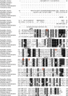
Sequence alignment of LaeA from A. nidulans with homologs in other fungal species. Amino acids with 80% or greater conservation are indicated in black. The arrow indicates the amino acid sequence missing at the N terminus of the LaeAΔ1–86 protein. Bars are used to denote the amino acids comprising the four common methyltransferase motifs of seven-β-strand methyltransferases. Red boxes highlight methionine residues mutated in this study. Amino acid sequences are taken from UniProt accession numbers C8VQG9, Q6TFC7, E0WDF6, G4XKY9, and I3RU94.
FIGURE 3.
FIGURE 3.
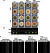
N-terminal 86 amino acids of LaeA are dispensable for function. Complementation of ΔlaeA was accomplished by targeted integration of constructs at the pyroA locus. A, strains of A. nidulans were grown for 4 days under light or dark conditions on glucose minimal media where null mutants of laeA produce fewer conidia and reduced colony pigmentation. As expected, a carboxyl GFP tag of laeA complements these gross phenotypes. Additionally, truncation of the N-terminal 86 amino acids (LaeAΔ1–86-GFP) also results in restoration of the gross phenotype of ΔlaeA strains. B, production of the mycotoxin sterigmatocystin was extracted and visualized via thin layer chromatography from cultures shown in A. Sterigmatocystin is nearly absent in ΔlaeA strains, and this phenotype is restored in both the LaeA-GFP and LaeAΔ1–86-GFP complementation strains. C, LaeA-GFP localized constitutively in the nucleus as expected, and the N-terminal truncation mutant LaeAΔ1–86-GFP also localizes exclusively in the nucleus. Histone H1 fused to mCherry (hhoA-mCherry) serves as a marker for nuclei. Strains used are as follows: WT (RJMP260.12), ΔlaeA (RJMP260.1), ΔlaeA laeA-GFP (RJMP261.26), and ΔlaeA laeAΔ1–86-GFP (RJMP264.5). DIC, differential interference contrast.
FIGURE 4.
FIGURE 4.
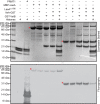
Recombinant LaeAΔ1–86 or MBP-LaeA does not methylate purified VeA, VelB, or histone proteins. In vitro methylation reactions were prepared with 1.7 μm [3H]AdoMet and incubated for 20 h at 37 °C with methyltransferases and potential methyl-accepting substrates, including 5 μg each of recombinant human histone H2A, H2B, H3.3, and H4, 1 μg of purified PRMT1 (positive control), 27.4 μg of MBP-LaeA, ∼5 μg of VeA, ∼5 μg of VelB, and 8.8 μg of LaeAΔ1–86. The samples were separated on a 10% BisTris gel with MES running buffer and stained with Coomassie. Polypeptide molecular weight markers (Bio-Rad broad range, ∼3 μg of each protein, catalogue number 161-0317) were electrophoresed in a parallel lane and shown on the left, and include myosin (200 kDa), β-galactosidase (116 kDa), phosphorylase b (97 kDa), serum albumin (66 kDa), ovalbumin (45 kDa), carbonic anhydrase (31 kDa), trypsin inhibitor (21 kDa), lysozyme (14 kDa), and aprotinin (6 kDa). Fluorography was performed by treating the gel with EN3HANCE and exposing the dried gels to film for 5 days at −80 °C as described under “Experimental Procedures.” Red asterisks indicate the position of MBP-LaeA and LaeAΔ1–86.
FIGURE 5.
FIGURE 5.
In vitro methylation assay using wild type, laeA overexpressing, and ΔlaeA nuclear extracts. Approximately 110 μg of crude nuclear protein from each strain was isolated as described under “Experimental Procedures” and incubated with 100 nm [3H]AdoMet for 4 h at 37 °C. When added, MBP-LaeA and LaeAΔ1–86 were present at 4 μg of protein. The samples were electrophoresed on a 10% Tris glycine polyacrylamide gel with Tris glycine running buffer and Coomassie-stained. Fluorography was performed by treating the gel with EN3HANCE and exposing the dried gel to film for 9 months at −80 °C. The positions of Bio-Rad broad range marker proteins are shown on the left. Recomb, recombinant.
FIGURE 6.
FIGURE 6.
In vitro methylation of wild type and ΔlaeA extracts of A. nidulans at vegetative, asexual, and sexual growth phases do not reveal LaeA methyl-accepting substrates. Whole cell extracts from wild type or ΔlaeA strains in vegetative (A), asexual (B), or sexual (C) growth stages were prepared as described under “Experimental Procedures.” Samples analyzed were as follows: lane 1, 300 μg of wild type whole cell extract; lane 2, 300 μg of ΔlaeA whole cell extract; lane 3, 300 μg of ΔlaeA whole cell extract mixed with 10 μg of purified MBP-LaeA, and lane 4, 300 μg of ΔlaeA whole cell extract mixed with 10 μg of purified LaeAΔ1–86. Each sample was incubated with 100 nm [3H]AdoMet for 3 h at 37 °C, separated by 10% Tris glycine polyacrylamide gels with Tris glycine running buffer, and stained with Coomassie. The gels were treated with EN3HANCE, and the dried gels were exposed to film for 1 month at −80 °C. The positions of Bio-Rad broad range marker proteins (M) are shown on the left. Red asterisks denote the position of MBP-LaeA or LaeAΔ1–86 in the Coomassie-stained gel and in the fluorograph.
FIGURE 7.
FIGURE 7.
In vivo protein methylation of wild type, laeA-overexpressing, and ΔlaeA A. nidulans strains separated into insoluble and soluble fractions. Aspergillus mycelia was labeled with [3H]AdoMet as described under “Experimental Procedures.” Lyophilized mycelia were ground to a powder and resuspended in 100 mm Tris-HCl, pH 7.5, 250 mm NaCl, 10% glycerol, 0.1% Nonidet P-40, and 1 mm EDTA. The resulting lysate was vortexed, kept at 0 °C for 10 min, and centrifuged at 20,000 × g for 20 min at 4 °C. The supernatant Soluble Fraction and the pellet Insoluble Fraction were fractionated by SDS-PAGE as described under “Experimental Procedures” on a 10% BisTris polyacrylamide gel with MES-SDS running buffer. A, Coomassie-stained gel. B, fluorograph of EN3HANCE-treated gels after 1 month at −80 °C. The positions of Bio-Rad broad range marker proteins are indicated.
FIGURE 8.
FIGURE 8.
A novel LaeA automethylated amino acid residue. 30 μg of purified LaeAΔ1–86 was incubated with 2.8 μm [3H]AdoMet for ∼20 h at 37 °C as described under “Experimental Procedures.” Reaction mixtures separated by 10% BisTris PAGE were stained, destained, and treated with EN3HANCE. After fluorography, the [3H]AdoMet-labeled LaeA protein band was excised, rehydrated with water, and acid-hydrolyzed as described under “Experimental Procedures.” After mixing with standards of methylated amino acids (1 μmol each) including 1-(π)-methylhistidine, ω-NG-monomethylarginine (ω-MMA), ω-NG,NG-dimethylarginine (ADMA), ω-NG,NG′-dimethylarginine (SDMA), ϵ-N-monomethyllysine hydrochloride (MMK), ϵ-N-dimethyllysine (DMK), and ϵ-N-trimethyllysine (TMK), high resolution cation exchange chromatography was performed as described under “Experimental Procedures.” The positions of the standard amino acids were determined by ninhydrin assay as described under “Experimental Procedures” and indicated by a dashed line. Radioactivity was determined in 200 μl of each fraction and is shown by a red solid line.
FIGURE 9.
FIGURE 9.
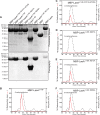
Direct demonstration of LaeA automethylation at methionine 207 by gel electrophoresis and high resolution cation exchange chromatography. Constructs were made to express His-tagged MBP fusion proteins of LaeAΔ1–86 where each methionine residue in the LaeAΔ1–86 sequence was replaced individually by an alanine residue. 150 μg of each of the purified LaeA proteins, with the exception of MBP-LaeA (lane 8, 82 μg), were incubated with 1.4 μm [3H]AdoMet for 20 h at 37 °C, and the reaction products were separated by 10% BisTris PAGE with MES running buffer as described under “Experimental Procedures.” A, gel was stained with Coomassie. The position of Bio-Rad broad range marker proteins are shown on the left. Controls included the unmutated MBP-LaeAΔ1–86 (2nd lane), unmutated LaeAΔ1–86 (5th lane), MBP alone (7th lane), and unmutated MBP-full-length LaeA (8th lane). Fluorography was performed by treating the gel with EN3HANCE and exposing the dried gel to film for 4 days at −80 °C. The portion of the EN3HANCE-treated gel containing the radiolabeled LaeA protein was excised, rehydrated with water, and acid-hydrolyzed as described under “Experimental Procedures.” B–F, after mixing with standards (1 μmol each, with the exception of S-methylmethionine with 5 μmol) of methylated amino acids, including 1-(π)-methylhistidine, S-methylmethionine, ω-NG-monomethylarginine, and ω-NG,NG-dimethylarginine, high resolution cation exchange chromatography was performed as described under “Experimental Procedures.” The positions of the standard amino acids were determined by ninhydrin assay as described under “Experimental Procedures” and indicated by a dashed line. Radioactivity was determined in 200 μl of each fraction and is shown in solid red lines.
FIGURE 10.
FIGURE 10.
OPA amino acid analysis verifies S-methylmethionine is the radioactive product of LaeA automethylation. Cation exchange chromatography fractions from the experiment shown in Fig. 9B were fluorescently labeled with OPA reagent and analyzed by reverse phase HPLC coupled to a fluorometer as described under “Experimental Procedures.” Fractions of 0.5 ml were collected, and radioactivity was quantified as described under “Experimental Procedures” (red line). The fluorescence HPLC trace (black line) represents relative fluorescence units (RFU); the OPA-derivative of S-methylmethionine elutes at 13.1 min.
FIGURE 11.
FIGURE 11.
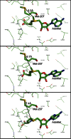
Modeling of the LaeA structure suggests that the sulfur atom of methionine 207 is close to the position of the transferable methyl group of bound AdoMet. The three-dimensional structure of full-length LaeA was based on the primary sequence (UniProt accession number C8VQG9) and derived from Phyre2 modeling; the position of the ligand AdoMet was modeled by 3DLigandSite. The positions of three possible conformations of AdoMet are shown that encompass the extent of the eight different ligand structures obtained. LaeA side chain residues within 4 Å of AdoMet are shown as thin lines with the exception of methionine 207, which is shown as a thick line with the sulfur atom in yellow. The bound AdoMet is also shown as a thick line. The distance of this sulfur atom to the transferable methyl group of AdoMet is 5.2, 6.7, and 7.4 Å in these three simulated conformations and is represented by black dashed lines.
FIGURE 12.
FIGURE 12.
Methionine 207 of LaeA is not required for function in vivo. A methionine 207 to alanine point mutation was constructed and integrated at the pyroA locus in a ΔlaeA strain of A. nidulans. A, LaeAM207A-GFP strains restored the gross phenotype of ΔlaeA strains as well as production of sterigmatocystin in both light and dark growth conditions. B, using fluorescent microscopy, LaeAM207A-GFP was found to be located in nuclei. C, quantification of asexual sporulation (conidiation) was measured on plates grown for 4 days under light conditions. Null mutants of laeA produce fewer conidia under these conditions. Complementation strains partially restore the production of conidia, although not to wild type levels. LaeA-GFP and LaeAM207A-GFP produce similar amounts of conidia. D, production of sexual spores (ascospores) is also reduced in ΔlaeA strains compared with wild type. This phenotype is partially restored in both LaeA-GFP and LaeAM207A-GFP complementation strains. Statistical differences were measured by analysis of variance using Prism 5.0 (GraphPad) and different letters represent statistical differences p < 0.001. Strains used in A, C, and D were WT (RJMP103.5), ΔlaeA (RJW41A), ΔlaeA laeA-GFP (RJMP256.3), and ΔlaeA laeAM207A-GFP (RJMP258.1). RJMP263.3 (ΔlaeA, hhoA-mCherry, pyroA::laeAM207A-GFP) was used in B.

Similar articles

Cited by

References

    1. Latgé J. P. (1999) Aspergillus fumigatus and aspergillosis. Clin. Microbiol. Rev. 12, 310–350 - PMC - PubMed
    1. Latgé J. P. (2001) The pathobiology of Aspergillus fumigatus. Trends Microbiol. 9, 382–389 - PubMed
    1. Brakhage A. A. (1998) Molecular regulation of β-lactam biosynthesis in filamentous fungi. Microbiol. Mol. Biol. Rev. 62, 547–585 - PMC - PubMed
    1. Alberts A. W., Chen J., Kuron G., Hunt V., Huff J., Hoffman C., Rothrock J., Lopez M., Joshua H., Harris E., Patchett A., Monaghan R., Currie S., Stapley E., Albers-Schonberg G., Hensens O., Hirshfield J., Hoogsteen K., Liesch J., Springer J. (1980) Mevinolin: a highly potent competitive inhibitor of hydroxymethylglutaryl-coenzyme A reductase and a cholesterol-lowering agent. Proc. Natl. Acad. Sci. U.S.A. 77, 3957–3961 - PMC - PubMed
    1. Keller N. P., Turner G., Bennett J. W. (2005) Fungal secondary metabolism–from biochemistry to genomics. Nat. Rev. Microbiol. 3, 937–947 - PubMed

Publication types

LinkOut - more resources