. 2013 May 10;288(19):13602-9.
doi: 10.1074/jbc.M112.443812. Epub 2013 Mar 26.

Structural and functional aspects of hetero-oligomers formed by the small heat shock proteins αB-crystallin and HSP27

Affiliations

Structural and functional aspects of hetero-oligomers formed by the small heat shock proteins αB-crystallin and HSP27

J Andrew Aquilina et al. J Biol Chem. .

Abstract

Background: αB-crystallin and HSP27 are mammalian intracellular small heat shock proteins.

Results: These proteins exchange subunits in a rapid and temperature-dependent manner.

Conclusion: This facile subunit exchange suggests that differential expression could be used by the cell to regulate the response to stress.

Significance: A robust technique defines parameters for the dynamic interaction between the major mammalian small heat shock proteins. Small heat shock proteins (sHSPs) exist as large polydisperse species in which there is constant dynamic subunit exchange between oligomeric and dissociated forms. Their primary role in vivo is to bind destabilized proteins and prevent their misfolding and aggregation. αB-Crystallin (αB) and HSP27 are the two most widely distributed and most studied sHSPs in the human body. They are coexpressed in different tissues, where they are known to associate with each other to form hetero-oligomeric complexes. In this study, we aimed to determine how these two sHSPs interact to form hetero-oligomers in vitro and whether, by doing so, there is an increase in their chaperone activity and stability compared with their homo-oligomeric forms. Our results demonstrate that HSP27 and αB formed polydisperse hetero-oligomers in vitro, which had an average molecular mass that was intermediate of each of the homo-oligomers and which were more thermostable than αB, but less so than HSP27. The hetero-oligomer chaperone function was found to be equivalent to that of αB, with each being significantly better in preventing the amorphous aggregation of α-lactalbumin and the amyloid fibril formation of α-synuclein in comparison with HSP27. Using mass spectrometry to monitor subunit exchange over time, we found that HSP27 and αB exchanged subunits 23% faster than the reported rate for HSP27 and αA and almost twice that for αA and αB. This represents the first quantitative evaluation of αB/HSP27 subunit exchange, and the results are discussed in the broader context of regulation of function and cellular proteostasis.

Keywords: Analytical Chemistry; Chaperone Chaperonin; Crystallin; HSP27; Mass Spectrometry (MS); Protein Aggregation; Small Heat Shock Proteins.


Figures
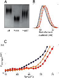
FIGURE 1.
FIGURE 1.
HSP27 and αB form hetero-oligomeric complexes in vitro. A, discontinuous native PAGE analysis of αB, HSP27, and an equimolar mixture of the two. The intermediate migration position of the mixture indicates that significant subunit exchange had occurred. B, DLS measurements demonstrated that, when mixed, αB (blue) and HSP27 (red) formed a polydisperse population (gold) with a size distribution intermediate between the two homo-oligomers. C, thermal denaturation curves for αB (blue), HSP27 (red), and an equimolar mixture of the two (gold). The change in average particle size (Z-average) as measured by DLS was used to estimate protein aggregation as the temperature was increased by 1 °C/min from 25 to 95 °C. Results shown are representative of three independent experiments.
FIGURE 2.
FIGURE 2.
Chaperone activity of hetero-oligomeric complexes formed between αB and HSP27. The ability of the homo- and hetero-oligomers to prevent the reduction-induced amorphous aggregation of bovine α-lactalbumin (A and B) and amyloid fibril formation of α-synuclein (C and D) was examined. Representative traces showing the change in light scatter at 360 nm (A) or ThT fluorescence emission at 490 nm (C) in the absence (purple) or presence of HSP27 (red), αB (blue), or an equimolar mixture of the two (gold) are shown. Molar ratios shown are 8:1 (α-lactalbumin:sHSPs) and 10:1 (α-synuclein:sHSPs). Samples containing αB (orange) or HSP27 (gray) alone overlap at the bottom of the trace. B and D, the percent protection afforded by the sHSPs at the various molar ratios (target protein:sHSPs) tested. Results shown are means ± S.E. of four independent experiments. *, significant (p < 0.05) difference compared with the αB and mixed oligomers at the same molar ratio.
FIGURE 3.
FIGURE 3.
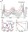
Dissociation mass spectrometry of HSP27 and αB. A, by careful selection of accelerating voltages across and argon gas flow into the collision cell, it was possible to remove two and three monomers from the hetero-oligomeric assemblies of HSP27 and αB, respectively. In the region of the spectrum assigned to the doubly stripped oligomers (n2), the peaks were sufficiently resolved such that assignments to individual oligomers could be made. B and C, expanded views of charge-state clusters on either side of the major overlapping peaks in the n2 region of αB and HSP27, respectively. Charge-state assignments were used to define the range of oligomers present in these sHSPs corresponding to the size exclusion chromatography peak maximum fractions.
FIGURE 4.
FIGURE 4.
Monitoring subunit exchange at 30 °C using mass spectrometry. A, the doubly stripped oligomeric region of the mass spectra for a mixture of HSP27 and αB was monitored over a 30-min period. A peak corresponding to the formation of a hetero-oligomer was observed to emerge at m/z ∼22,400, accompanied by a minor decay in the relative intensities of the homo-oligomeric signature peaks at m/z ∼20,160 (αB), and m/z ∼22,700 (HSP27). The genesis and progression of the hetero-oligomeric peak indicate that exchange occurred predominantly via incorporation of αB subunits into HSP27 oligomers. The 30-min time point spectrum is colored for contrast. B, monitoring the reaction for a sufficient period of time demonstrated that there was a complete exchange of subunits between HSP27 and αB. The spectra are colored to distinguish overlapping traces. C, subunit exchange kinetics of HSP27 and αB as a function of temperature. Logarithmic plots of homo-oligomeric decay versus time at 37 °C (red circles), 30 °C (yellow squares) and 23 °C (blue diamonds), demonstrating a linear relationship and therefore a first order-type reaction. Rate constants for subunit exchange at each temperature were determined from the slope of the corresponding plot.

Similar articles

Cited by

References

    1. Dobson C. M. (2003) Protein folding and misfolding. Nature 426, 884–890 - PubMed
    1. Ecroyd H., Carver J. A. (2009) Crystallin proteins and amyloid fibrils. Cell. Mol. Life Sci. 66, 62–81 - PMC - PubMed
    1. Chiti F., Dobson C. M. (2006) Protein misfolding, functional amyloid, and human disease. Annu. Rev. Biochem. 75, 333–366 - PubMed
    1. Dobson C. M. (2001) The structural basis of protein folding and its links with human disease. Philos. Trans. R. Soc. Lond. B Biol. Sci. 356, 133–145 - PMC - PubMed
    1. Muchowski P. J. (2002) Protein misfolding, amyloid formation, and neurodegeneration: a critical role for molecular chaperones? Neuron 35, 9–12 - PubMed

Publication types