. 2013 May 3;288(18):12605-14.
doi: 10.1074/jbc.M112.399477. Epub 2013 Mar 26.

Smoothened oligomerization/higher order clustering in lipid rafts is essential for high Hedgehog activity transduction

Affiliations

Smoothened oligomerization/higher order clustering in lipid rafts is essential for high Hedgehog activity transduction

Dawei Shi et al. J Biol Chem. .

Abstract

The Hedgehog (Hh) signaling pathway plays evolutionarily conserved roles in controlling embryonic development and tissue homeostasis, and its dysregulation has been implicated in many human diseases including congenital disorder and cancer. The Hh pathway has a unique signal reception system that includes two membrane proteins, the receptor Patched (Ptc) and the transducer Smoothened (Smo). In the Hh signaling cascade, Smo plays a critical role in controlling transduction of Hh gradient signal from the outside into the inside of cells. Although the Smo downstream signal transduction has been intensively studied, the mechanism by which Smo on the plasma membrane is regulated has not been fully understood. As a specific membrane structure of metazoan cells, lipid rafts act as a platform to regulate signal transduction by forming a nanoscale cluster through protein-protein or protein-lipid interactions. However, it remains largely unknown whether lipid rafts are also involved in the regulation of Hh signal transduction. Here, we show that Smo extracellular domain (N terminus) and transmembrane domains form oligomers/higher order clusters in response to Hh signal. Furthermore, we identify that lipid rafts on the plasma membrane are essential for high level activity of Smo during the Hh signal transduction. Finally, our observation suggests that oligomerization/higher order clustering of Smo C-terminal cytoplasmic tail (C-tail) is essential for the transduction of high level Hh signal. Collectively, our data support that in response to Hh gradient signals, Smo transduces high level Hh signal by forming oligomers/higher order clusters in the lipid rafts of cell plasma membrane.

Keywords: Cancer; Drosophila; Hedgehog; Lipid Raft; Oligomer; Signal Transduction; Smo.


Figures
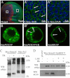
FIGURE 1.
FIGURE 1.
Smo extracellular domains form oligomers/higher order clusters in the presence of Hh. A–A″, endogenous Smo forms clusters in the presence of Hh. The wild-type Drosophila wing disc was stained with anti-Smo and anti-Ci antibodies to label Smo (green) and Ci (red), and DAPI was used to mark nucleus (blue). A, Ci staining positive part (red) or negative part (red signal negative) delimits the A compartment, in which there is no Hh, and the P compartment, in which there is high level Hh. The squares delimit the magnified areas in A′ and A″. The image magnified from the P compartment (A′) or from the A compartment (A″) shows Smo and DAPI staining. Scale bars, 50 μm (A) or 5 μm (A′ and A″). B–B″, S2 cells were transfected with the indicated constructs and observed under a Leica Confocal microscope. B, SmoWTCFP locates in the cytoplasm in the absence of Hh. B′ and B″, SmoWTCFP in the presence of Hh or SmoSDCFP accumulates on the plasma membrane and forms clusters. Arrows point the clustered Smo in A′, B′, and B″. C, to detect the oligomers of SmoΔC, a native gel electrophoresis was employed. To compare the ratio between oligomer and monomer of SmoΔC with or without Hh treatment, the loading amount of the first two samples was adjusted to make the total SmoΔC to a comparable level. Western blotting shows that, upon Hh treatment, SmoΔC forms oligomers; meanwhile, the level of SmoΔC monomer decreases. As the control, the level of Fz2 monomer, dimer, or oligomer does not change with or without Hh treatment. Arrows point to the monomer, and arrowheads point to the dimer and oligomer, respectively. D, in Western blots, the autophosphorylated tyrosine level of Myc-SmoΔC-EphB2CT was detected by 4G10 antibody. In the presence of Hh, the tyrosine phosphorylation level of Myc-SmoΔC-EphB2CT is increased. Tyrosine phosphorylation could be erased after lysates were treated with LAR, a protein-tyrosine phosphatase.
FIGURE 2.
FIGURE 2.
The proximity of Smo intracellular subdomains is induced by Hh. A, S2 cells were transfected with indicated constructs and treated with or without Hh, respectively. CFP and YFP signals were acquired for FRET before (BP) and after (AP) photobleaching YFP at the top half of each cell (white rectangle frame). B, FRET efficiency is shown from the indicated CFP/YFP-tagged constructs, which were transfected into S2 cells and treated with or without Hh (mean ± S.D. (error bars), n ≥ 30; *, p < 0.05). C, diagram shows the Smo truncations which were used in D and E. D, S2 cells were co-transfected with Myc-tagged or FLAG-tagged full-length Smo (SmoFL) or Smo truncations, respectively. The immunoprecipitation was followed with mouse anti-FLAG antibody, and Western blotting (WB) was followed with rabbit anti-FLAG or anti-Myc antibodies for immunoprecipitation product and cell lysates. Full-length and truncated Smo, beside SmoCT, interact with each other. E, S2 cells were transfected with the indicated constructs, and then native gel electrophoresis was employed. Western blotting shows that full-length and truncated Smo, beside SmoCT, form dimer or oligomer. Arrows point to monomer, and arrowheads point to dimer and oligomer.
FIGURE 3.
FIGURE 3.
Lipid rafts are important for Smo function. A, S2 cells were co-transfected with Myc-tagged Smo and HA-tagged dGq and then treated with or without Hh, respectively. Membrane lipid microdomains were fractionated with a three-step sucrose gradient centrifugation. Myc-tagged Smo and HA-tagged dGq were detected by Western blotting. Myc-Smo or HA-dGq in cell lysates shows the indicated protein expression level. B, ptc-Luc reporter assay of S2 cells transfected with the indicated constructs and treated with or without Hh, PUFA, or SA is shown (mean ± S.D. (error bars), triplicate wells; *, p < 0.05; **, p < 0.01). C and D, to detect FRET efficiency, S2 cells were transfected with the indicated CFP/YFP-tagged constructs and treated with or without Hh, SA, or PUFA, respectively. C, graph shows that PUFA treatment reduces the FRET of SmoCFPC/SmoYFPC and SmoCFPL3/SmoYFPL3 (mean ± S.D., n ≥ 30; *, p < 0.05' **, p < 0.01). D, CFP and YFP signals from SmoCFPL3/SmoYFPL3 in the absence/presence of Hh were acquired for FRET before (BP) and after (AP) photobleaching YFP (for details of FRET, see “Experimental Procedures” and legend of Fig. 2A). E–H″, cell surface accumulation of CFP-tagged Smo treated with (F–H″) or without (E–E″) Hh, PUFA (G–G″), or SA (H–H″). S2 cells were transfected with CFP-tagged Smo followed by immunostaining with anti-SmoN antibody before membrane permeabilization. Cell surface accumulated Smo (red) and total Smo (green, CFP) are shown. Arrows in F and H point to Smo clusters on the plasma membrane. The images were taken from videos (please refer to supplemental Videos 1–4).
FIGURE 4.
FIGURE 4.
Smo locates in lipid rafts in vivo in the presence of Hh. FLAG-tagged SmoWT was expressed in the A compartment with Ci-Gal4 (A–A‴ and B–B‴), or in the P compartment with Hh-Gal4 (C–C‴ and D–D‴). FITC-CTB (green) was employed to mark lipid rafts on the plasma membrane. Anti-FLAG antibody was used to immunostain the overexpressed Smo (red), and DAPI was used to mark nucleus (blue). The squares in A–A‴ and C–C‴ delimit the magnified areas in B–B‴ and D–D‴, respectively. The magnified images showed the details of Smo, CTB, and DAPI staining (B–B‴ and D–D‴). In the absence of Hh, Smo located in cytoplasm and could not co-localize with CTB (B and B′). In the presence of Hh, however, Smo co-localized with CTB on the plasma membrane (D and D′). Scale bars, 50 μm (A‴ and C‴) or 5 μm (B‴ and D‴).
FIGURE 5.
FIGURE 5.
Lipids rafts are essential for high level Hh signal transduction. Wing imaginal discs of third instar larvae expressing FLAG-tagged SmoSD (A–F′) or HA-tagged full-length Ci (G–H′) with MS1096;ptc-LacZ are shown. Discs were dissected and treated with SA (served as control, A–A‴, C–C‴, E–E′, and G–G′) or PUFA (B–B‴, D–D‴, F–F′, and H–H′) in serum-free M3 medium for 6 h and then immunostained with different antibodies to show the expression of ptc-LacZ (blue), En (blue), Ci (red), and SmoSD (green). E–E′ and F–F′ are the magnified views of FLAG staining to show the clusters on the plasma membrane of SmoSD. DAPI staining labeled the nucleus. Arrows in E point to the oligomers/clusters of SmoSD on the cell surface. Scale bars, 5 μm (E′ and F′). G–H′, wild-type Ci was overexpressed with MS1096 to induce En expression in the A compartment. PUFA treatment did not reduce En expression level (G and H). I, S2 cells were transfected with Myc-SmoΔC and treated with SA or PUFA in the absence/presence of Hh. Cell lysates were harvested for native gel electrophoresis and then detected by anti-Myc antibody in Western blotting (WB). J, graph shows the ratio change of oligomer/monomer in the experiment.
FIGURE 6.
FIGURE 6.
Oligomerization/higher order clustering of Smo C-tail is essential for high activity of Hh pathway. A, upper left, amino acid sequences of CCm, CCd, and CCt, with green and blue letters indicating different amino acids among them. Upper right, diagrams of Myr-FLAG-CCm-SmoCT, Myr-FLAG-CCd-SmoCT, and Myr-FLAG-CCt-SmoCT. The lower diagram shows Myr-FLAG-CC-SmoCT variants. B–M″, wing discs dissected from third instar larvae, which were expressing the indicated Smo C-tail mutants (E–M″) or wild-type fly (B–D″) with MS1096;dpp-LacZ, were immunostained to show the expression level of dpp-LacZ (blue), Ptc (blue), En (blue), Ci (red) and FLAG (green), respectively.

Similar articles

Cited by

References

    1. Ingham P. W., McMahon A. P. (2001) Hedgehog signaling in animal development: paradigms and principles. Genes Dev. 15, 3059–3087 - PubMed
    1. Ingham P. W., Nakano Y., Seger C. (2011) Mechanisms and functions of Hedgehog signalling across the metazoa. Nat. Rev. 12, 393–406 - PubMed
    1. Jiang J., Hui C. C. (2008) Hedgehog signaling in development and cancer. Dev. Cell 15, 801–812 - PMC - PubMed
    1. Wilson C. W., Chuang P. T. (2010) Mechanism and evolution of cytosolic Hedgehog signal transduction. Development 137, 2079–2094 - PMC - PubMed
    1. Hooper J. E., Scott M. P. (1989) The Drosophila patched gene encodes a putative membrane protein required for segmental patterning. Cell 59, 751–765 - PubMed

Publication types

MeSH terms

LinkOut - more resources