. 2013 Mar 26;4(2):e00636-12.
doi: 10.1128/mBio.00636-12.

Inhibition of viral pathogenesis and promotion of the septic shock response to bacterial infection by IRF-3 are regulated by the acetylation and phosphorylation of its coactivators

Affiliations

Inhibition of viral pathogenesis and promotion of the septic shock response to bacterial infection by IRF-3 are regulated by the acetylation and phosphorylation of its coactivators

Saurabh Chattopadhyay et al. mBio. .

Abstract

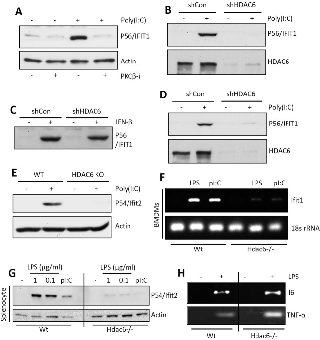
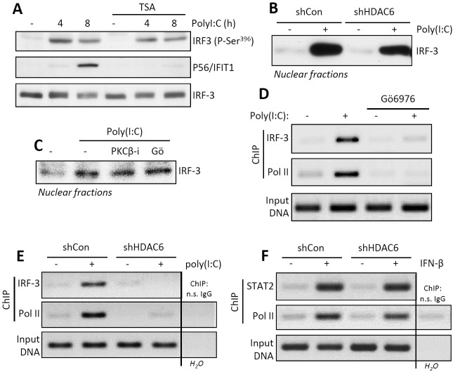
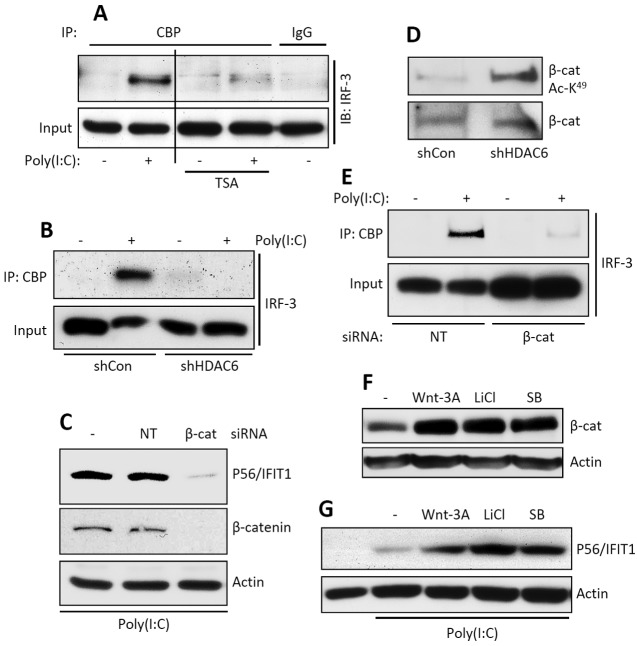
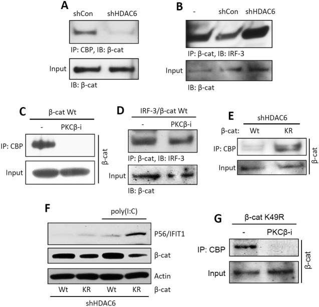
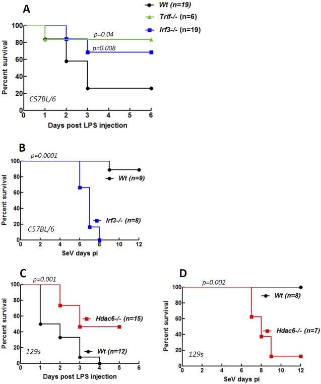
Interferon (IFN) is required for protecting mice from viral pathogenesis; reciprocally, it mediates the deleterious septic shock response to bacterial infection. The critical transcription factor for IFN induction, in both cases, is IRF-3, which is activated by TLR3 or RIG-I signaling in response to virus infection and TLR4 signaling in response to bacterial infection. Here, we report that IRF-3's transcriptional activity required its coactivators, β-catenin and CBP, to be modified by HDAC6-mediated deacetylation and protein kinase C isozyme β (PKC-β)-mediated phosphorylation, respectively, so that activated nuclear IRF-3 could form a stable transcription initiation complex at the target gene promoters. β-Catenin bridges IRF-3 and CBP, and the modifications were required specifically for the interaction between β-catenin and CBP but not β-catenin and IRF-3. Consequently, like IRF-3(-/-) mice, HDAC6(-/-) mice were resistant to bacterial lipopolysaccharide-induced septic shock. Conversely, they were highly susceptible to pathogenesis caused by Sendai virus infection. Thus, HDAC6 is an essential component of the innate immune response to microbial infection.


Figures

FIG 1
FIG 1
PKC-β and HDAC6 are required for TLR-induced IRF-3 transcriptional activity. (A) P56/IFIT1 induction was analyzed in TLR3-stimulated HT1080 cells in the presence of a PKC-β inhibitor by Western blotting. (B) P56/IFIT1 induction was analyzed in TLR3 [poly(I⋅C), 100 µg/ml]-stimulated shCon and shHDAC6 cells by Western blotting. (C) P56/IFIT1 induction was analyzed in IFN-β (1,000 U/ml)-stimulated shCon or shHDAC6 cells by Western blotting. (D) P56/IFIT1 induction was analyzed in RLH [transfected-poly(I⋅C)]-stimulated shCon or shHDAC6 cells by Western blotting. (E) P54/Ifit2 induction was analyzed in RLH [transfected-poly(I⋅C)]-stimulated wt or HDAC6−/− MEFs by Western blotting. KO, knockout. (F) Primary bone marrow-derived macrophages (BMDMs) from wt or Hdac6−/− mice were treated with TLR4 (LPS, 1 µg/ml) or TLR3 [poly(I⋅C) (pI:C), 25 µg/ml] agonists, Ifit1 induction was analyzed by RT-PCR, and 18S rRNA was used as a loading control. (G) Splenocytes isolated from wt or Hdac6−/− mice were treated with LPS (at the indicated concentrations) or poly(I⋅C) (50 µg/ml), and P54/Ifit2 induction was analyzed by Western blotting. (H) BMDMs were isolated from wt or Hdac6−/− mice, and IL-6 (Il6) and TNF-α induction was analyzed by RT-PCR upon LPS treatment.
FIG 2
FIG 2
PKC-β and HDAC6 activities are required for promoter occupancy of IRF-3 but not its phosphorylation and nuclear translocation. (A) IRF-3 phosphorylation (P-Ser396) and P56/IFIT1 induction in TLR3-stimulated HT1080 cells in the absence or the presence of TSA. (B) Nuclear translocation of IRF-3 was analyzed in TLR3-stimulated shCon or shHDAC6 cells by Western blotting. (C) Nuclear translocation of IRF-3 was analyzed in PKC-β inhibitor- or Gӧ6976-treated, TLR3-stimulated HT1080 cells by Western blotting. (D, E) Chromatin immunoprecipitation (ChIP) of IRF-3 and RNA Pol II on the IFIT1 promoter was analyzed in TLR3-stimulated, Gӧ6976-treated HT1080 (D) and shHDAC6 (E) cells. (F) ChIP of STAT2 and RNA Pol II on the IFIT1 promoter was analyzed in IFN-β-stimulated shCon and shHDAC6 cells. n.s., nonspecific.
FIG 3
FIG 3
HDAC6-deficient cells fail to recruit complex containing IRF-3, CBP, and β-catenin. (A) Co-IP of IRF-3 with CBP in TLR3-stimulated HT1080 cells in the absence or the presence of TSA. The input represents the levels of IRF-3 in cell lysates. IB, immunoblotting. (B) Co-IP of IRF-3 with CBP in TLR3-stimulated shCon and shHDAC6 cells. The input represents the levels of IRF-3 in cell lysates. (C) HT1080 cells were transfected with nontargeting (NT) or β-catenin-specific siRNA, and induction of P56/IFIT1 was analyzed upon TLR3 stimulation by Western blotting. Knockdown of β-catenin was confirmed by Western blotting (middle panel). (D) β-Catenin acetylation (acetylated K49 [Ac-K49]-specific antibody) was analyzed in shCon and shHDAC6 cells by Western blotting. (E) Co-IP of IRF-3 with CBP in nontargeting (NT) or β-catenin (β-cat)-specific, siRNA-transfected HT1080 cells upon TLR3 stimulation. The input represents levels of β-catenin in cell lysates. (F, G) Western analyses of β-catenin levels (F) and P56/IFIT1 induction (G) in TLR3-stimulated [poly(I⋅C)] HT1080 cells in the absence or the presence of Wnt signaling activators (as indicated). Actin was used as a loading control. SB, SB216763.
FIG 4
FIG 4
HDAC6-mediated deacetylated β-catenin is required for interaction with CBP but not IRF-3. (A) Flag-tagged β-catenin was expressed in shCon and shHDAC6 cells. IP of β-catenin with CBP was analyzed upon TLR3 stimulation. The input represents the levels of β-catenin in the cell lysates. (B) Flag-tagged β-catenin and V5-tagged IRF-3 were coexpressed in shCon and shHDAC6 cells. Co-IP of β-catenin with IRF-3 was analyzed upon TLR3 stimulation. The input represents the levels of β-catenin in the cell lysates. (C) Flag-tagged β-catenin was expressed in HT1080 cells. Co-IP of β-catenin with CBP was analyzed upon TLR3 stimulation in the absence or the presence of the PKC-β inhibitor (PKCβ-i). The input represents the levels of β-catenin in the cell lysates. (D) Flag-tagged β-catenin and V5-tagged IRF-3 were coexpressed in HT1080 cells. Co-IP of β-catenin with IRF-3 was analyzed upon TLR3 stimulation in the absence or the presence of the PKC-β inhibitor. The input represents the levels of β-catenin in the cell lysates. (E) Flag-tagged β-catenin (wt or the K49R mutant [KR]) was expressed in shHDAC6 cells. Co-IP of β-catenin with CBP was analyzed upon TLR3 stimulation. The input represents the levels of β-catenin in the cell lysates. (F) The wt or K49R mutant of β-catenin was expressed in shHDAC6 cells, and P56/IFIT1 induction was analyzed upon TLR3 stimulation [poly(I⋅C)]. (G) The Flag-tagged β-catenin mutant (K49R) was expressed in HT1080 cells. Co-IP of β-catenin with CBP was analyzed upon TLR3 stimulation in the absence or the presence of the PKC-β inhibitor. The input represents the levels of β-catenin in the cell lysates.
FIG 5
FIG 5
HDAC6-mediated transcriptional activation of IRF-3 contributes to LPS-induced septic shock and protection against Sendai virus infection in mice. Shown are percentages of survival of IRF3−/−, TRIF−/−, and wt mice (C57BL/6) after intraperitoneal injection with LPS (1 mg/mouse) (A), of IRF3−/− and wt mice (C57BL/6) after intranasal infection with SeV (strain 52, 1,000 PFU/mouse) (B), of HDAC6−/− and wt mice (129s) after intraperitoneal injection with LPS (1 mg/mouse) (C), and of HDAC6−/− and wt mice (129s) after intranasal infection with SeV (strain 52, 1,000 PFU/mouse). The statistical significance of survival differences between the knockout mice and their wt controls are indicated as P values. pi, postinfection.

References

    1. Fensterl V, Sen GC. 2009. Interferons and viral infections. Biofactors 35:14–20 - PubMed
    1. Borden EC, Sen GC, Uze G, Silverman RH, Ransohoff RM, Foster GR, Stark GR. 2007. Interferons at age 50: past, current and future impact on biomedicine. Nat. Rev. Drug Discov. 6:975–990 - PMC - PubMed
    1. Nagarajan U. 2011. Induction and function of IFNβ during viral and bacterial infection. Crit. Rev. Immunol. 31:459–474 - PMC - PubMed
    1. Schoggins JW, Rice CM. 2011. Interferon-stimulated genes and their antiviral effector functions. Curr. Opin. Virol. 1:519–525 - PMC - PubMed
    1. Karaghiosoff M, Steinborn R, Kovarik P, Kriegshäuser G, Baccarini M, Donabauer B, Reichart U, Kolbe T, Bogdan C, Leanderson T, Levy D, Decker T, Müller M. 2003. Central role for type I interferons and Tyk2 in lipopolysaccharide-induced endotoxin shock. Nat. Immunol. 4:471–477 - PubMed

Publication types

Associated data