

doi: 10.1002/anie.201208987.
Epub 2013 Mar 26.
The flavoprotein dodecin as a redox probe for electron transfer through DNA
Affiliations
- PMID: 23532984
- PMCID: PMC3743158
- DOI: 10.1002/anie.201208987
Item in Clipboard
The flavoprotein dodecin as a redox probe for electron transfer through DNA
Angew Chem Int Ed Engl.
.
Free PMC article
No abstract available
Figures
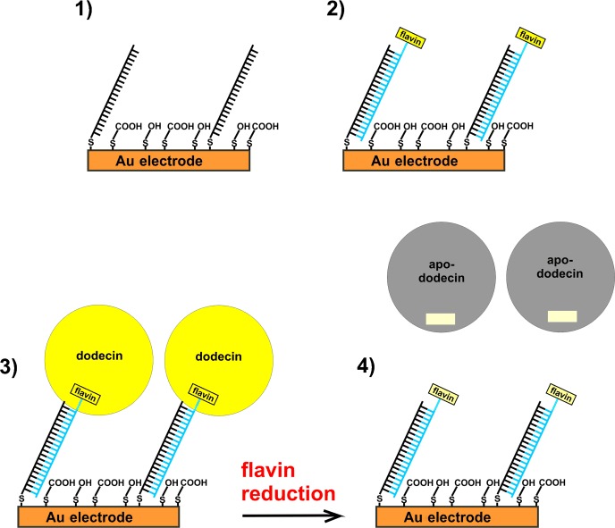
Experimental approach using dodecin to probe electron-transfer through DNA. 1) A monolayer of disulfide modified ss-DNA is adsorbed on gold whereupon a mixture of mercaptobutanol (MCB) and mercaptopropionic acid (MPA) in a ratio of 1:1 is added to release non-specifically adsorbed DNA and to increase the hybridization efficiency. 2) Complementary flavin-modified ss-DNA is hybridized. 3) Dodecin is reconstituted at the surface. 4) A negative potential is applied. If electron transfer through ds-DNA is possible, this results in flavin reduction followed by the release of apododecin (only one of the six flavin binding pockets of dodecin is shown).
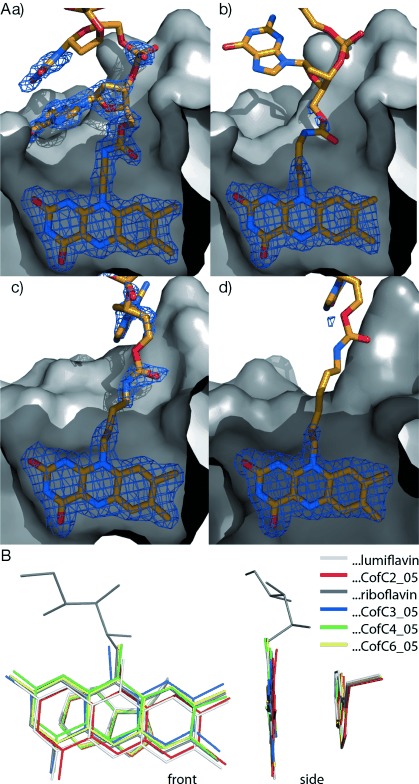
X-ray structures of CofCn_O5 flavin–DNA ligands in the tE dodecin binding pocket. Dodecin is depicted as apoprotein in a surface representation (gray) with the incorporated ligands shown in stick representation (C orange, N blue, O red). Dodecin has six binding pockets for the incorporation of dimers of flavins in a C2-symmetric manner. For clarity the C2-symmetric part of one binding pocket is shown. Structural data used for figures are deposited with the protein data bank (pdb) codes 2vkg, 2vkf, and 4b2h (http://www.rcsb.org ). A) tE dodecin with bound CofCn_O5 ligands with varying lengths of alkyl chain connecting isoalloxazine to DNA. CofC2_O5 (a) has an ethyl chain, CofC3_O5 (b) propyl, CofC4_O5 (c) butyl, and CofC6_O5 (d) hexyl. Electron omit density is shown at σ=1.5 as a blue mesh, highlighting protein-bound substructures of ligands. The unbound parts of the ligands, which could not be identified by electron density, are shown for clarity in one possible conformation. (B) Superposition of Cα-backbone-aligned tE dodecins with CofCn_O5 ligands (and tryptophan, which is part of the binding site) in front and side view. Ligands are depicted by their isoalloxazine anchor. Wildtype dodecin with riboflavin and lumiflavin ligands are shown for comparison.
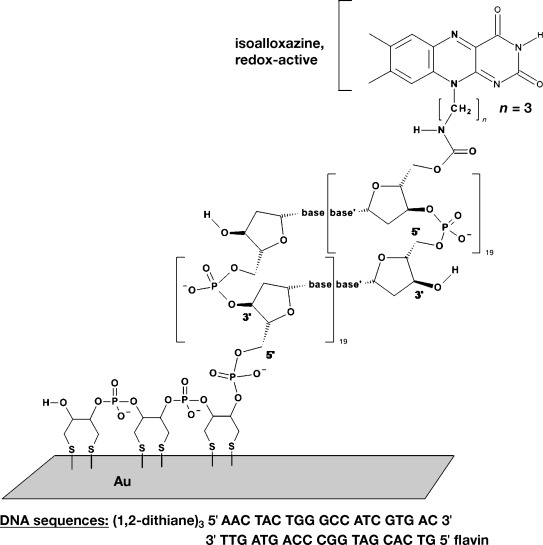
Structure and sequence of the flavin- and disulfide-modified DNA.
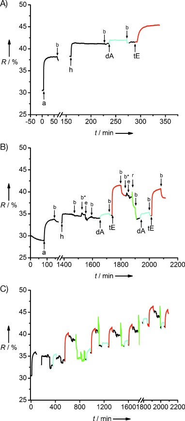
Kinetic SPR scan curves A) adsorption of disulfide modified ss-DNA (a), rinsing with buffer (b), hybridization with flavin-modified ss-DNA (h), incubation of non-binding apododecin as negative control (dA), reconstitution of dodecin (tE). During addition of MCB/MPA in water the reflectivity R changed to values below 10 %, because of the difference in reflective index between water and buffer (not shown). B) The experimental procedure was extended by the following steps: rinsing with argon saturated buffer (b*), electrochemistry, that is, application of −550 mV vs. Ag/AgCl for 5 min then cyclic voltammetry (e), chemical reduction using sodium dithionite in argon-saturated buffer (r). C) Long-term run. Incubation of dA and rinsing with buffer is shown in cyan, incubation of tE and rinsing with buffer is shown red, reduction and rinsing is shown in green.
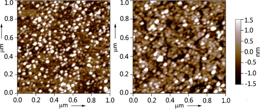
AFM images (acquired in air) of the dodecin reconstituted on the flavin terminated ds-DNA monolayer adsorbed on template-stripped gold prepared using the same concentrations for DNA adsorption and hybridization as for SPR measurements (left) and lower flavin concentration (right).
References
-
- Ito T, Rokita SE. Angew. Chem. 2004;116:1875–1878.
-
- Angew. Chem. Int. Ed. 2004;43:1839–1842. - PubMed
-
- Ito T, Rokita SE. J. Am. Chem. Soc. 2003;125:11480–11481. - PubMed
-
- Fazio D, Trindler C, Heil K, Chatgilialoglu C, Carell T. Chem. Eur. J. 2011;17:206–212. - PubMed
-
- Behrens C, Burgdorf LT, Schwogler A, Carell T. Angew. Chem. 2002;114:1841–1844. - PubMed
Publication types
MeSH terms
Substances
LinkOut - more resources
Full Text Sources
Other Literature Sources
