Comparative Study
. 2013 May;92(5):444-9.
doi: 10.1177/0022034513483315. Epub 2013 Mar 26.

Type I interferon receptor deficiency prevents murine Sjogren's syndrome

Affiliations
Comparative Study

Type I interferon receptor deficiency prevents murine Sjogren's syndrome

B M Szczerba et al. J Dent Res. 2013 May.

Abstract

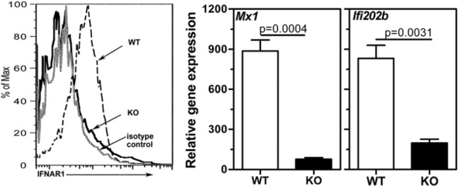
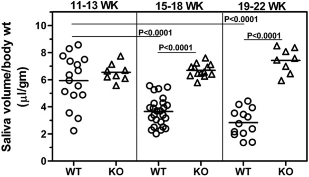
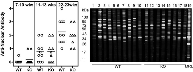
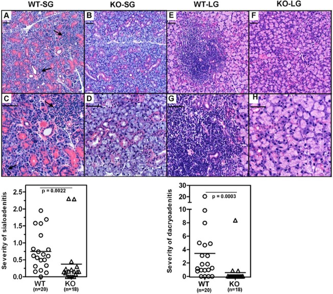
In Sjögren's Syndrome (SS), inherent glandular defects, autoimmunity, and mononuclear cell infiltration within the salivary glands cause reduced salivation leading to xerostomia. Excessive production of type I interferons (IFN), triggered by environmental and genetic factors, is considered pathogenic in this disorder. However, whether type I IFN production is causative or an outcome of the disease process is not known. To address this question, we introduced a deficiency of interferon alpha receptor 1 (Ifnar1) into B6.Aec1Aec2 mice, which are known to have the genetic loci necessary for developing a SS-like disorder. This new mouse strain, B6.Aec1Aec2Ifnar1 (-/-), lacking type I IFN-mediated signaling, was characterized for pilocarpine-induced salivation, the presence of serum autoantibodies, sialoadenitis, and dacryoadenitis. Compared with the B6.Aec1Aec2Ifnar1 (+/+) (wild-type) mice, the B6.Aec1Aec2Ifnar1 (-/-) (knockout) mice had significantly lower mononuclear cell infiltration in the salivary and lacrimal glands. The knockout mice were completely protected from salivary gland dysfunction. Surprisingly, they had a robust autoantibody response comparable with that of the wild-type mice. These findings demonstrate that, in the absence of type I IFN-mediated signaling, systemic autoantibody responses can be dissociated from glandular pathology. Our study suggests that, in genetically susceptible individuals, the type I IFN pathway can instigate certain features of SS.


Conflict of interest statement

The authors declare no potential conflicts of interest with respect to the authorship and/or publication of this article.

Figures

Figure 1.
Figure 1.
Lack of IFNAR in B6Aec1Aec2 mice suppresses the expression of type I IFN-responsive genes Mx1 and Ifi202b. (Left panel) Flow cytometry analysis showing a representative histogram of IFNAR staining on PBMCs obtained from WT (dotted line) and KO (solid line) mice. The staining by isotype control is shown by a gray line. (Right panel) Gene expression levels of Mx1 and Ifi202b in the submandibular glands obtained from WT (n = 4) and KO (n = 3) mice at 8 wks of age were determined by real-time PCR with Taqman primers. The data are represented as mean ± SEM relative gene expression over a pooled RNA sample from an age-matched C57BL/6 mouse, which was used as the calibrator. Gapdh was used to normalize the expression. Statistical significance was determined by the Mann-Whitney test. Similar results were obtained in an additional experiment.
Figure 2.
Figure 2.
Saliva production decreases over time in WT mice but not in KO mice that lack the type I IFN signaling pathway. Pilocarpine-induced saliva production was measured at the different ages indicated in the Fig. Data are represented as the ratio of saliva volume (µL) to body weight (gm). Student’s t test was used to determine statistical significance. Saliva volumes (mean ± SEM) in WT mice at 15 to 18 wks (3.65 ± 0.21) and 19 to 22 wks (2.83 ±0.28) were significantly lower compared with those at 11 to 13 wks (5.93 ± 0.46) of age, indicating a progressive loss of function. In contrast, in KO mice, the mean saliva volumes between early and late ages were not different. Although WT and KO mice had comparable saliva production at 11 to 13 wks of age, the differences between the 2 groups were highly significant at the 15 to 18-week (p < .0001) and 19- to 22-week (p < .0001) time-points.
Figure 3.
Figure 3.
Both WT and KO mice generated a robust autoantibody response. Sera obtained from WT and KO mice at different time-points were analyzed for the presence of autoantibodies. (Left panel) Intensity of ANA staining by indirect immunofluorescence. Representative ANA pictures are shown in Appendix Fig. 3. The incidence and severity of ANA staining were not different between the WT and KO mice. (Right panel) A representative image of a Western blot showing reactivity to proteins from a mouse submandibular gland cell line. All sera were used at 1:200 dilutions, and goat anti-mouse IgG antibody coupled to IRDye 800CW was used to detect bound antibodies. Lanes 1 to 10 are sera from individual WT mice, lanes 11 to 18 are sera from individual KO mice, and lane 19 has pooled sera from lupus-prone MRL lpr/lpr mice. Similar results were obtained in an additional experiment.
Figure 4.
Figure 4.
The IFNAR deficiency suppresses mononuclear cell infiltration within the submandibular and lacrimal glands. Submandibular (A-D) and lacrimal glands (E-H) obtained from WT (n = 20) and KO (n = 18) mice were evaluated for mononuclear cell infiltration in H&E-stained sections. The inflammation was quantitated by an individual blinded to experimental details. A, B, E, and F show representative low-magnification images, and C, D, G, and H are images at higher magnification. Severity of sialoadenitis and dacryoadenitis (lower panels) represents the fraction of area covered by inflammatory infiltrates in salivary and lacrimal glands, respectively, for each mouse. Statistical significance was calculated by the Mann-Whitney test. The mean severity scores of sialoadenitis and dacryoadenitis in the WT mice were significantly higher than those in the KO mice. The arrows indicate the foci of mononuclear cell infiltration. Scale bar = 50 microns.

References

    1. Agrawal H, Jacob N, Carreras E, Bajana S, Putterman C, Turner S, et al. (2009). Deficiency of type I IFN receptor in lupus-prone New Zealand mixed 2328 mice decreases dendritic cell numbers and activation and protects from disease. J Immunol 183:6021-6029. - PMC - PubMed
    1. Bagavant H, Thompson C, Ohno K, Setiady Y, Tung KS. (2002). Differential effect of neonatal thymectomy on systemic and organ-specific autoimmune disease. Int Immunol 14:1397-1406. - PubMed
    1. Carr AJ, Ng WF, Figueiredo F, Macleod RI, Greenwood M, Staines K. (2012). Sjögren’s syndrome— an update for dental practitioners. Br Dent J 213:353-357. - PubMed
    1. Cha S, Nagashima H, Brown VB, Peck AB, Humphreys-Beher MG. (2002). Two NOD Idd associated intervals contribute synergistically to the development of autoimmune exocrinopathy (Sjögren’s syndrome) on a healthy murine background. Arthritis Rheum 46:1390-1398. - PubMed
    1. Delaleu N, Nguyen CQ, Peck AB, Jonsson R. (2011). Sjögren’s syndrome: studying the disease in mice. Arthritis Res Ther 13:217. - PMC - PubMed

Publication types

MeSH terms

Substances

LinkOut - more resources