

Surface plasmon resonance: a useful technique for cell biologists to characterize biomolecular interactions
- PMID: 23533209
- PMCID: PMC3608497
- DOI: 10.1091/mbc.E12-10-0713
Surface plasmon resonance: a useful technique for cell biologists to characterize biomolecular interactions
Abstract
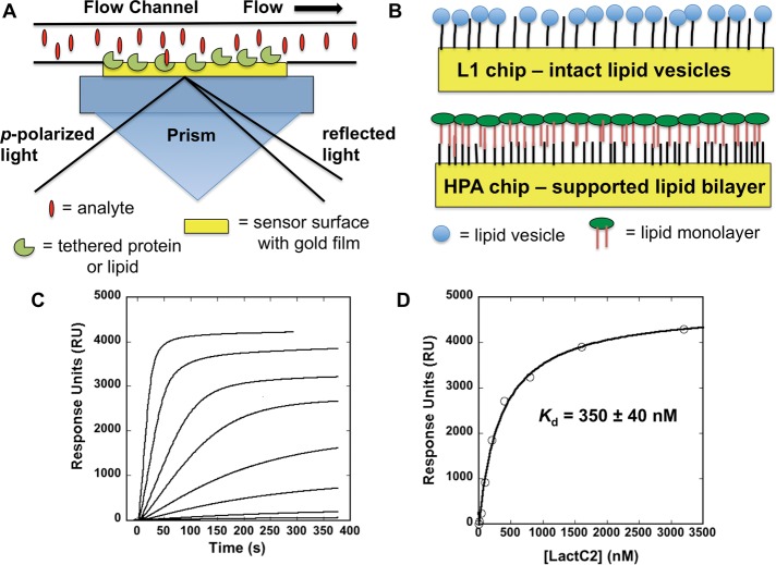
Surface plasmon resonance (SPR) is a powerful technique for monitoring the affinity and selectivity of biomolecular interactions. SPR allows for analysis of association and dissociation rate constants and modeling of biomolecular interaction kinetics, as well as for equilibrium binding analysis and ligand specificity studies. SPR has received much use and improved precision in classifying protein-protein interactions, as well as in studying small-molecule ligand binding to receptors; however, lipid-protein interactions have been underserved in this regard. With the field of lipids perhaps the next frontier in cellular research, SPR is a highly advantageous technique for cell biologists, as newly identified proteins that associate with cellular membranes can be screened rapidly and robustly for lipid specificity and membrane affinity. This technical perspective discusses the conditions needed to achieve success with lipid-protein interactions and highlights the unique lipid-protein interaction mechanisms that have been elucidated using SPR. It is intended to provide the reader a framework for quantitative and confident conclusions from SPR analysis of lipid-protein interactions.
Figures
Similar articles
-
Surface plasmon resonance for measuring interactions of proteins with lipid membranes.Methods Mol Biol. 2013;974:23-36. doi: 10.1007/978-1-62703-275-9_2. Methods Mol Biol. 2013. PMID: 23404270
-
Surface plasmon resonance in protein-membrane interactions.Chem Phys Lipids. 2006 Jun;141(1-2):169-78. doi: 10.1016/j.chemphyslip.2006.02.010. Epub 2006 Mar 20. Chem Phys Lipids. 2006. PMID: 16584720 Review.
-
Quantitative analysis of molecular partition towards lipid membranes using surface plasmon resonance.Sci Rep. 2017 Mar 30;7:45647. doi: 10.1038/srep45647. Sci Rep. 2017. PMID: 28358389 Free PMC article.
-
Using Surface Plasmon Resonance to Quantitatively Assess Lipid-Protein Interactions.Methods Mol Biol. 2016;1376:141-53. doi: 10.1007/978-1-4939-3170-5_12. Methods Mol Biol. 2016. PMID: 26552681 Free PMC article.
-
Peeking into a secret world of pore-forming toxins: membrane binding processes studied by surface plasmon resonance.Toxicon. 2003 Sep;42(3):225-8. doi: 10.1016/s0041-0101(03)00197-1. Toxicon. 2003. PMID: 14559072 Review.
Cited by
-
Sensitivity Analysis of Single- and Bimetallic Surface Plasmon Resonance Biosensors.Sensors (Basel). 2021 Jun 25;21(13):4348. doi: 10.3390/s21134348. Sensors (Basel). 2021. PMID: 34202104 Free PMC article.
-
A Library Screening Strategy Combining the Concepts of MS Binding Assays and Affinity Selection Mass Spectrometry.Front Chem. 2019 Oct 4;7:665. doi: 10.3389/fchem.2019.00665. eCollection 2019. Front Chem. 2019. PMID: 31637233 Free PMC article.
-
Lysosomal integral membrane protein-2 as a phospholipid receptor revealed by biophysical and cellular studies.Nat Commun. 2017 Dec 4;8(1):1908. doi: 10.1038/s41467-017-02044-8. Nat Commun. 2017. PMID: 29199275 Free PMC article.
-
Peptides derived from MARCKS block coagulation complex assembly on phosphatidylserine.Sci Rep. 2017 Jun 27;7(1):4275. doi: 10.1038/s41598-017-04494-y. Sci Rep. 2017. PMID: 28655899 Free PMC article.
-
Navigating the Expansive Landscapes of Soft Materials: A User Guide for High-Throughput Workflows.ACS Polym Au. 2023 Dec 5;3(6):406-427. doi: 10.1021/acspolymersau.3c00025. eCollection 2023 Dec 13. ACS Polym Au. 2023. PMID: 38107416 Free PMC article. Review.
References
-
- Bigay J, Antonny B. Curvature, lipid packing, and electrostatics of membrane organelles: defining cellular territories in determining specificity. Dev Cell. 2012;23:886–895. - PubMed
-
- Cho W, Stahelin RV. Membrane-protein interactions in cell signaling and membrane trafficking. Annu Rev Biophys Biomol Struct. 2005;34:119–151. - PubMed
-
- Erb EM, Chen X, Allen S, Roberts CJ, Tendler SJ, Davies MC, Forsen S. Characterization of the surface generated by liposome binding to the modified dextran matrix of a surface plasmon resonance sensor chip. Anal Biochem. 2000;280:29–35. - PubMed
-
- Höning S, Ricotta D, Krauss M, Spate K, Spolaore B, Motley A, Robinson M, Robinson C, Haucke V, Owen DJ. Phosphatidylinositol-(4,5)-bisphosphate regulates sorting signal recognition by the clathrin-associated adaptor complex AP2. Mol Cell. 2005;18:519–531. - PubMed
-
- Kretschmann E, Raether H. Radiative decay of non-radiative surface plasmons excited by light. Z Naturforsch. 1968;230:2135–2136.
Publication types
MeSH terms
Substances
Grants and funding
LinkOut - more resources
Full Text Sources
Other Literature Sources
