Clinical Trial
. 2013 May;3(5):534-47.
doi: 10.1158/2159-8290.CD-12-0502. Epub 2013 Mar 26.

De-repression of PDGFRβ transcription promotes acquired resistance to EGFR tyrosine kinase inhibitors in glioblastoma patients

Affiliations
Clinical Trial

De-repression of PDGFRβ transcription promotes acquired resistance to EGFR tyrosine kinase inhibitors in glioblastoma patients

David Akhavan et al. Cancer Discov. 2013 May.

Abstract

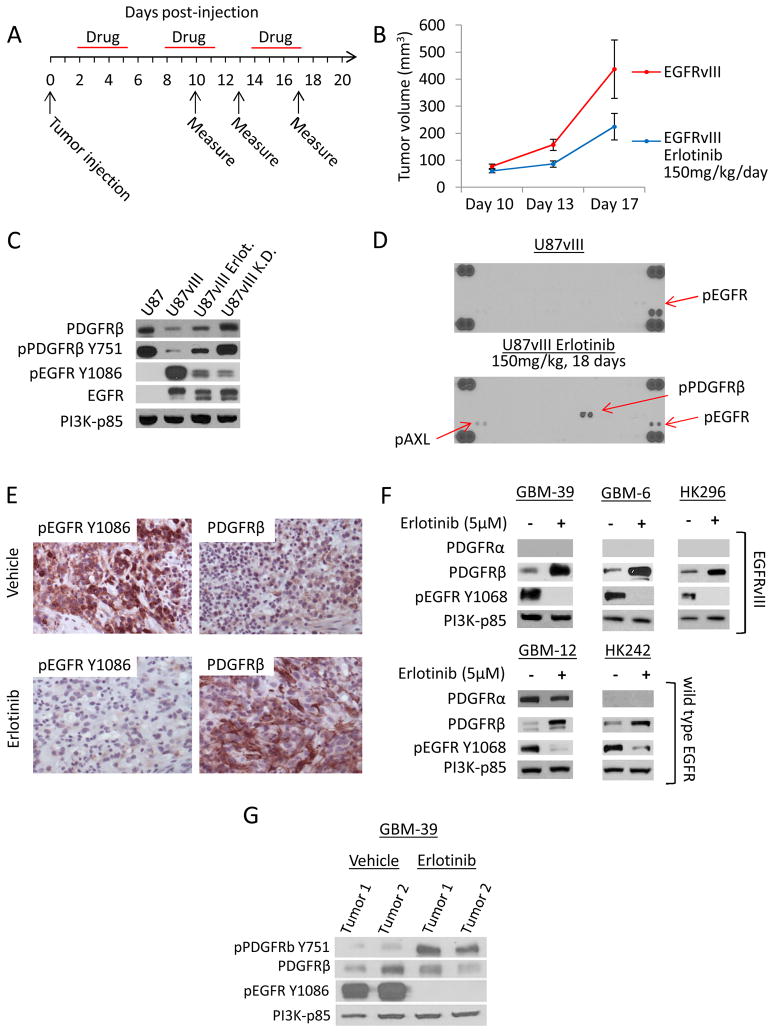
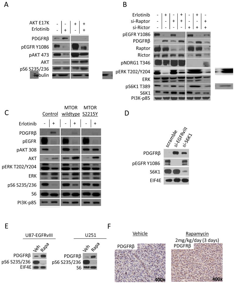
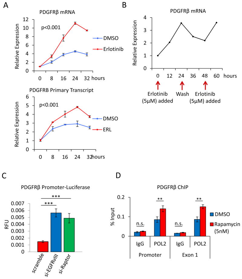
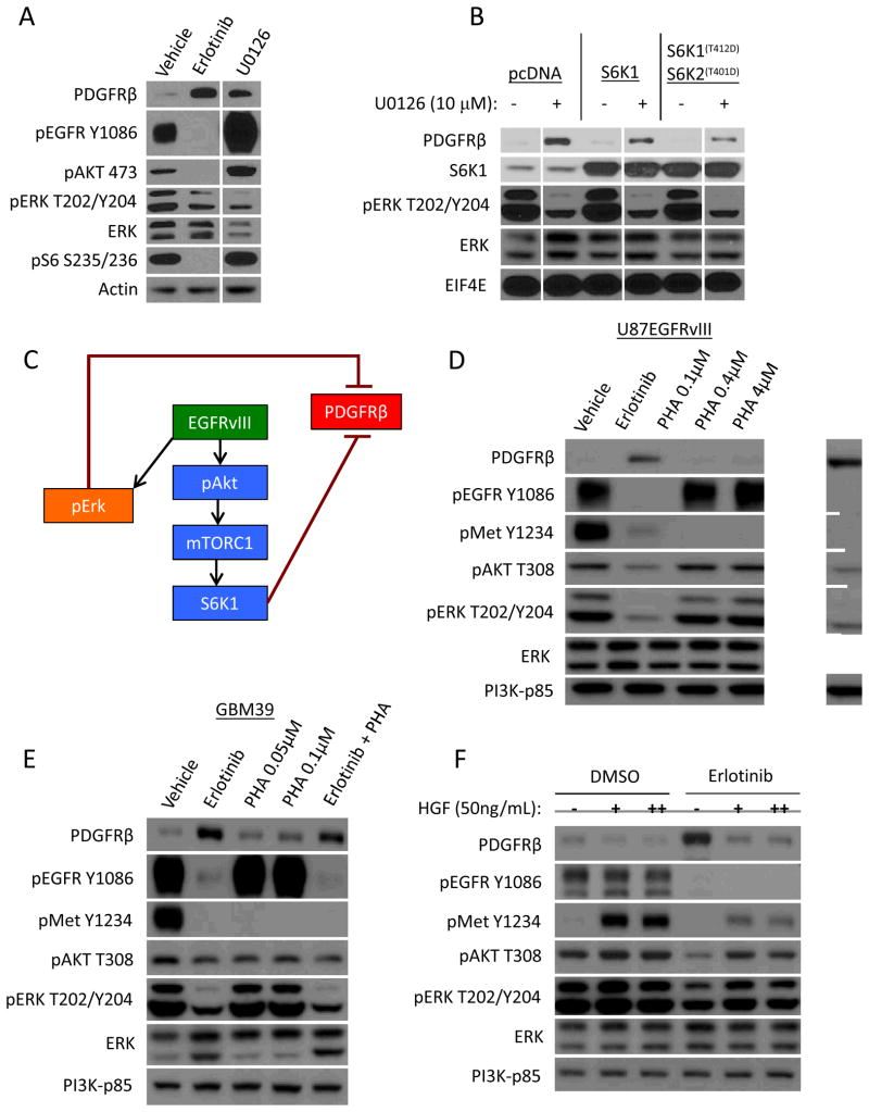
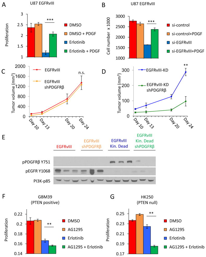
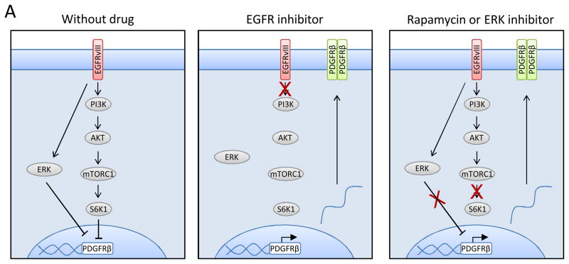
Acquired resistance to tyrosine kinase inhibitors (TKI) represents a major challenge for personalized cancer therapy. Multiple genetic mechanisms of acquired TKI resistance have been identified in several types of human cancer. However, the possibility that cancer cells may also evade treatment by co-opting physiologically regulated receptors has not been addressed. Here, we show the first example of this alternate mechanism in brain tumors by showing that EGF receptor (EGFR)-mutant glioblastomas (GBMs) evade EGFR TKIs by transcriptionally de-repressing platelet-derived growth factor receptor β (PDGFRβ). Mechanistic studies show that EGFRvIII signaling actively suppresses PDGFRβ transcription in an mTORC1- and extracellular signal-regulated kinase-dependent manner. Genetic or pharmacologic inhibition of oncogenic EGFR renders GBMs dependent on the consequently de-repressed PDGFRβ signaling for growth and survival. Importantly, combined inhibition of EGFR and PDGFRβ signaling potently suppresses tumor growth in vivo. These data identify a novel, nongenetic TKI resistance mechanism in brain tumors and provide compelling rationale for combination therapy.

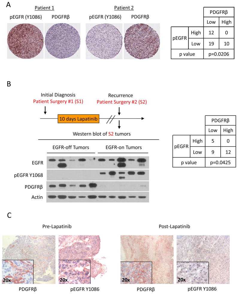
Significance: These results provide the fi rst clinical and biologic evidence for receptor tyrosinekinase (RTK) "switching" as a mechanism of resistance to EGFR inhibitors in GBM and provide a molecular explanation of how tumors can become "addicted" to a non amplified, nonmutated, physiologically regulated RTK to evade targeted treatment.


Conflict of interest statement

Conflict of interest disclosure statement:

Drs. Mischel and Cloughesy served on an advisory board for Celgene’s mTOR kinase inhibitor program. Dr. Mischel and Cloughesy also collaborated with Celgene and Sanofi through research contracts on their mTOR kinase, and PI3K/mTOR kinase inhibitor clinical trials. Dr. Kornblum collaborated with Celgene on a research contract for the mTOR kinase inhibitor program. The authors are not aware of any other potential conflicts of interest.

Figures

Fig 1
Fig 1. A reciprocal relationship between EGFR and PDGFRβ in glioma
(A) Experimental design of a mouse model of EGFR inhibitor resistance. U87-EGFRvIII cells were subcutaneously implanted in the mouse flank on day zero. Mice were treated with erlotinib (150mg/kg) on day two and as indicated thereafter. (B) Tumor growth curve of U87-EGFRvIII xenografts in mice treated with erlotinib (150mg/kg as indicated in (A)) or vehicle. (C) Immunoblot of indicated tumor lysates determining total and phospho-PDGFRβ or EGFR from U87, U87-EGFRvIII +/− eroltinib treatment and U87-EGFRvIII kinase dead xenografts harvested on day 21. PI3K-p85 is used as a loading control in this and subsequent immunoblots. (D) Receptor Tyrosine Kinase (RTK) array of 42 RTKs performed on U87-EGFRvIII xenograft lysates on day 21 from mice treated with erlotinib or vehicle as described in (A). (E) IHC of PDGFRβ and phospho-EGFR in vehicle and erlotinib-treated U87-EGFRvIII orthotopic xenografts. (F) Immunoblot of PDGFRα, PDGFRβ and phospho-EGFR from patient-derived GBM neurospheres expressing EGFRvIII or wildtype EGFR as indicated. Whole cell lysates were collected after 24 hours of erlotinib or vehicle treatment. (G) Immunoblot of tumor lysates from EGFRvIII expressing GBM-39 xenografts following oral gavage with vehicle or erlotinib for ten days.
Fig. 2
Fig. 2. PDGFRβ expression is suppressed in EGFR activated GBMs
(A) IHC staining for phospho-EGFR and PDGFRβ in clinical GBM tissues. The p value indicated was calculated using Fisher’s exact test. (B) Immunoblot of PDGFRβ and EGFR in clinical GBM tumors samples treated with lapatinib. Patients were treated with lapatinib for ten days following initial diagnosis. Second tumor samples were obtained following recurrence. Tumor lysates were prepared and grouped according to phospho-EGFR status (EGFR-off/on). The p value was calculated using Fisher’s exact test. (C) IHC staining for PDGFRβ and phospho-EGFR in pre and post lapatinib-treated GBM tissue.
Fig. 3
Fig. 3. EGFRvIII suppresses PDGFRβ through AKT and mTORC1 signaling
(A) Immunoblot of PDGFRβ and indicated proteins in U87-EGFRvIII cells expressing constitutively active AKT1 (E17K) treated with erlotinib (5μM) for 24 h. (B) Immunoblot of lysates from U87-EGFRvIII cells with transient knockdown of MTOR complex proteins Raptor or Rictor and treated with erlotinib (5μM) as indicated. (C) Immunoblot of U87-EGFRvIII cells expressing constitutively active (S2215Y) or wildtype mTOR and treated with erlotinib (5μM) for 24 h as indicated. (D) Immunoblot of PDGFRβ levels in response to transient knockdown of EGFRvIII, or S6 kinase 1 in U87-EGFRvIII cells. (E) PDGFRβ levels in U87-EGFRvIII and U251 cells treated with vehicle or rapamycin (5nM) for 24 hours. (F) IHC of PDGFRβ in intracranial U251 GBM tumors following 3 days of rapamycin (2 mg/kg/day) or vehicle treatment.
Fig. 4
Fig. 4. EGFR signaling regulates transcription of PDGFRβ gene
(A) Time course of PDGFRβ primary transcript and mRNA expression in U87-EGFRvIII cells treated with erlotinib (5μM) or vehicle for up to 32 h. (B) Determination of PDGFRβ mRNA expression in response to erlotinib treatment or media washout over 60 h. (C) Luciferase assay comparing PDGFRβ promoter activity in U87-EGFRvIII cells transfected with scrambled siRNA to siRNA against raptor or EGFRvIII. (D) Chromatin immunoprecipitation of RNA Polymerase II at the promoter or exon 1 of PDGFRβ gene following treatment with vehicle or rapamycin (5nM) for 24 h. *p<0.05, **p<0.01, ***p<0.001.
Fig. 5
Fig. 5. ERK signaling contributes to the regulation of PDGFRβ
(A) Immunoblot of PDGFRβ and indicated proteins from whole cell lysates of U87-EGFRvIII cells treated with MEK inhibitor U0126 (5μM), erlotinib (5μM) or vehicle for 24 h. (B) Determination of PDGFRβ protein levels from U87-EGFRvIII cells transfected with empty vector (pcDNA), wild type S6K1, or constitutively active S6K1 (T412D) and S6K2 (T401D). In addition cells were treated with MEK inhibitor U0126 (10μM) or vehicle for 24 h as indicated. (C) A schematic of the signals downstream of EGFRvIII regulating PDGFRβ protein expression. (D, E) PDGFRβ protein levels from U87-EGFRvIII cells (D) or patient derived neurosphere GBM-39 (E) treated with erlotinib (5μM) or MET inhibitor PHA at the indicated dose. (F) Immunoblot of PDGFRβ and indicated proteins from U87-EGFRvIII cells treated with erlotinib and MET ligand HGF (50–100 ng/mL) for 24 h as indicated.
Fig. 6
Fig. 6. PDGFRβ is dispensable for EGFRvIII-driven GBM growth, but is required for the optimal growth of EGFR-inhibited tumors
(A) Proliferation of U87-EGFRvIII cells over 4 days treated with erlotinib (5μM) or PDGF-bb ligand (20ng/mL) alone or in combination as indicated. Eroltinib was added on day zero of culture and PDGF-bb was added daily at 20ng/mL thereafter. (B) Growth of U87-EGFRvIII cells transiently transfected with control scrambled siRNA or siEGFRvIII and treated with PDGF-bb as described in (A). (C, D) Growth curve of xenografts subcutaneously implanted with U87EGFRvIII, U87EGFRvIII/shPDGFRβ, U87EGFRvIII kinase dead, or U87EGFRvIII kinase dead/shPDGFRβ cells as indicated. (E) Immunoblot of phospho-PDGFRβ and EGFR from lysates harvested on day 24 from tumors as described in C and D. (F, G) Proliferation of EGFRvIII expressing patient-derived neurospheres GBM-39 and HK-250 treated with erlotinib (5μm) and PDGFRβ inhibitor AG1295 (3μM) alone or in combination as indicated. *p<0.05, **p<0.01, ***p<0.001.
Fig. 7
Fig. 7. Model of proposed RTK-switch
Under conditions of heightened growth receptor signaling (e.g., EGFRvIII mutation), PDGFRb expression is repressed by downstream ERK and mTOR activity. Inhibition of these growth pathways, such as EGFR- or mTOR-inhibitors results in the transcription of the PDGFRβ gene and the upregulation of PDGFβ receptor.

Similar articles

Cited by

References

    1. Paez JG, Janne PA, Lee JC, Tracy S, Greulich H, Gabriel S, et al. EGFR mutations in lung cancer: correlation with clinical response to gefitinib therapy. Science. 2004;304:1497–500. - PubMed
    1. Mellinghoff IK, Wang MY, Vivanco I, Haas-Kogan DA, Zhu S, Dia EQ, et al. Molecular determinants of the response of glioblastomas to EGFR kinase inhibitors. N Engl J Med. 2005;353:2012–24. - PubMed
    1. Ang KK, Berkey BA, Tu X, Zhang HZ, Katz R, Hammond EH, et al. Impact of epidermal growth factor receptor expression on survival and pattern of relapse in patients with advanced head and neck carcinoma. Cancer Res. 2002;62:7350–6. - PubMed
    1. Pao W, Chmielecki J. Rational, biologically based treatment of EGFR-mutant non-small-cell lung cancer. Nat Rev Cancer. 2010;10:760–74. - PMC - PubMed
    1. Sequist LV, Waltman BA, Dias-Santagata D, Digumarthy S, Turke AB, Fidias P, et al. Genotypic and histological evolution of lung cancers acquiring resistance to EGFR inhibitors. Sci Transl Med. 2011;3:75ra26. - PMC - PubMed

Publication types

MeSH terms