. 2013 Apr 9;110(15):5957-62.
doi: 10.1073/pnas.1219232110. Epub 2013 Mar 26.

Paradoxical activation and RAF inhibitor resistance of BRAF protein kinase fusions characterizing pediatric astrocytomas

Affiliations

Paradoxical activation and RAF inhibitor resistance of BRAF protein kinase fusions characterizing pediatric astrocytomas

Angela J Sievert et al. Proc Natl Acad Sci U S A. .

Erratum in

  • Proc Natl Acad Sci U S A. 2013 May 21;110(21):8750

Abstract

Astrocytomas are the most common type of brain tumors in children. Activated BRAF protein kinase mutations are characteristic of pediatric astrocytomas with KIAA1549-BRAF fusion genes typifying low-grade astrocytomas and (V600E)BRAF alterations characterizing distinct or higher-grade tumors. Recently, BRAF-targeted therapies, such as vemurafenib, have shown great promise in treating V600E-dependent melanomas. Like (V600E)BRAF, BRAF fusion kinases activate MAPK signaling and are sufficient for malignant transformation; however, here we characterized the distinct mechanisms of action of KIAA1549-BRAF and its differential responsiveness to PLX4720, a first-generation BRAF inhibitor and research analog of vemurafenib. We found that in cells expressing KIAA1549-BRAF, the fusion kinase functions as a homodimer that is resistant to PLX4720 and accordingly is associated with CRAF-independent paradoxical activation of MAPK signaling. Mutagenesis studies demonstrated that KIAA1549-BRAF fusion-mediated signaling is diminished with disruption of the BRAF kinase dimer interface. In addition, the KIAA1549-BRAF fusion displays increased binding affinity to kinase suppressor of RAS (KSR), an RAF relative recently demonstrated to facilitate MEK phosphorylation by BRAF. Despite its resistance to PLX4720, the KIAA1549-BRAF fusion is responsive to a second-generation selective BRAF inhibitor that, unlike vemurafenib, does not induce activation of wild-type BRAF. Our data support the development of targeted treatment paradigms for BRAF-altered pediatric astrocytomas and also demonstrate that therapies must be tailored to the specific mutational context and distinct mechanisms of action of the mutant kinase.


Conflict of interest statement

The authors declare no conflict of interest.

Figures

Fig. 1.
Fig. 1.
Cells expressing KIAA1549-BRAF fusions display resistance and an associated enhanced paradoxical activation in response to first generation targeted BRAF inhibition. (A) KIAA1549-BRAF fusions are the result of an internal tandem duplication event that fuses KIAA1549, an uncharacterized gene, with the C terminus BRAF kinase domain (lacking the autoinhibitory N terminus). Constructs used in this study include Fusion-1 (long-form fusion protein), Fusion-2 (short-form fusion protein), and Fusion-3 and Fusion-4, proteins further truncated to eliminate putative KIAA1549 transmembrane domains, with Fusion-3 further modified to include an N terminus Src myristoylation sequence for membrane localization. Wild-type BRAF and V600EBRAF sequences are shown for comparison. CR1, conserved region 1; CR2, conserved region 2; CR3, conserved region 3; CRD, cystine-rich domain; RBD, RAS-binding domain. (B) BRAF fusion constructs demonstrate similar responses to targeted BRAF inhibition with PLX4720 in in vitro kinase assays. Myc-tagged V600EBRAF and KIAA1549-BRAF Fusion-1–Fusion-4 were transiently overexpressed in HEK293T cells. Kinases were immunoprecipitated, and in vitro kinase assays were performed with purified MEK in the presence of increasing concentrations of PLX4720 (0, 0.1,1, and 10 µM) over 30 min. The inhibitory effects of increasing concentrations of PLX4720 were evaluated by Western blot analysis using anti–phospho-MEK1/2. (C) Cell lines stably overexpressing the BRAF fusion constructs demonstrate resistance and enhanced paradoxical activation in the presence of targeted BRAF inhibition with PLX4720. The Western blots show MAPK pathway responsiveness in the presence of increasing concentrations (0, 0.01, 0.1, 1, and 10 µM) of PLX4720 in stably selected NIH/3T3 KIAA1549-BRAF cell lines (Left), Ba/F3 KIAA1549-BRAF cell lines (Center), and murine cortical neurosphere KIAA1549-BRAF cell lines (Right). As described previously for neurospheres, V600EBRAF-expressing cells entered senescence (26) and thus were excluded from this analysis. (D) NIH/3T3 cells stably expressing KIAA1549-BRAF Fusion-1–Fusion-4 display increased anchorage-independent cell growth in the presence of increasing concentrations of PLX4720 (0, 0.1, 1, and 10 µM) compared with cells expressing wild-type BRAF. (E) NIH/3T3 cells stably expressing KIAA1549-BRAF Fusion-3 (Upper) and Fusion-4 (Lower) cells demonstrate accelerated tumor growth in the presence of PLX4720. On the day of injection, mice were started on treatment with PLX4720-infused chow or received control chow. Both fusion cell lines were resistant to PLX4720. Data are mean ± SEM of five mice. The y axis shows tumor volume in cubic millimeters; x axis, days since injection. (F) Ba/F3 cells stably expressing KIAA-BRAF Fusion-4 demonstrate increased cell proliferation and accelerated tumor growth in the presence of PLX4720. (Upper) The IL-3–independent stably selected Ba/F3 Fusion-4 cell line demonstrates increased cell proliferation in the presence of PLX4720 (0.1 µM for 2 wk). Data are percent change in mean cell count ± SEM. (Lower) The Ba/F3 KIAA1549-BRAF Fusion-4 cell line was injected into the flanks of balb/c nu/nu mice. On the day of injection, mice were started on treatment with PLX4720-infused chow or received control chow. Results are comparable to those found with the NIH/3T3 KIAA1549-BRAF fusion cell lines, with the Ba/F3 Fusion-4 tumors demonstrating resistance to PLX4720. Data are mean ± SEM of five mice. The y axis shows tumor volume in cubic millimeters; the x axis, days since injection.
Fig. 2.
Fig. 2.
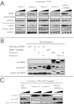
BRAF fusions function as distinct, altered signaling complexes. (A) CRAF is dispensable for drug resistance and associated paradoxical activation in KIAA1549-BRAF fusion-expressing cells. Using RNA interference, CRAF was knocked down in NIH/3T3 BRAF cell lines stably expressing KIAA1549-BRAF fusions, and the effects of increasing concentrations of PLX4720 (0, 0.1, 1, and 10 µM) were evaluated by Western blot analysis. No change in paradoxical activation as evaluated by pMEK immunoblotting was seen in the NIH/3T3 cells expressing fusion constructs. (B) KIAA1549-BRAF fusions signal as constitutive homodimers. To compare levels of kinase dimerization, GST pull-downs were performed from NIH/3T3 cells combinatorially coexpressing Myc-tagged and GST-tagged wild-type BRAF, V600EBRAF, and indicated KIAA1549-BRAF fusions. Western blot analysis with anti-Myc or anti-GST antibodies was performed as indicated. Robust fusion–fusion homodimerization is evident with no evidence for fusion heterodimerization with wild-type BRAF or homodimerization of V600EBRAF. G, GST-tagged constructs; M, Myc-tagged constructs. (C) KIAA1549-BRAF fusions display enhanced interactions with KSR1. Stably selected NIH/3T3 KIAA1549-BRAF cell lines expressing Myc-tagged wild-type BRAF, Fusion-3, Fusion-4, and V600EBRAF constructs were transfected with GST-KSR1 and incubated with increasing concentrations of PLX4720 (0, 1, and 10 µM). GST pull-downs were performed, and protein interactions were assessed by Western blot analysis. KSR-1 displayed enhanced affinity for Fusion-3 and Fusion-4 compared with wild-type BRAF or V600EBRAF.
Fig. 3.
Fig. 3.
BRAF fusion signaling is impaired by disruption of the dimerization interface. (A) Dimerization interface mutation impairs BRAF fusion homodimerization. To compare levels of kinase dimerization, GST pull-downs were performed from lysates of NIH/3T3 cells combinatorially coexpressing Myc-tagged and GST-tagged constructs. Robust fusion–fusion homodimerization was evident, but this dimerization was impaired when the R509H mutant was coexpressed. G, GST-tagged constructs; M, Myc-tagged constructs. (B) R509H mutant BRAF fusion constructs fail to demonstrate enhanced activation in response to PLX4720. MEK phosphorylation was assessed in NIH/3T3 cells stably expressing KIAA1549-BRAF fusions and the corresponding R509H mutants after incubation with increasing concentrations (0, 0.1, 1, and 10 µM) of PLX4720 for 30 min. (C) NIH/3T3 cells stably expressing KIAA1549-BRAF fusion R509H mutants display reduced transforming potential, associated with decreased anchorage-independent cell growth, and do not exhibit enhanced growth in the presence of increasing concentrations of PLX4720 (0, 0.1, 1, and 10 µM). (D) Disruption of the dimerization interface with a R509H mutation results in impaired tumor growth in vivo. NIH/3T3 cells stably expressing KIAA1549-BRAF fusions or the corresponding R509H mutants were injected into the flank of balb/c nu/nu mice. Data are mean ± SEM of five mice. The y axis indicates tumor volume in cubic millimeters; the x axis, days since injection.
Fig. 4.
Fig. 4.
BRAF fusion targeting by second-generation BRAF inhibitors. (A) BRAF fusion constructs demonstrate similar responses to selective BRAF inhibition with PLX PB-3 in in vitro kinase assays. Myc-tagged V600EBRAF and KIAA1549-BRAF Fusion 1–Fusion 4 were transiently overexpressed in HEK293T cells. Kinases were immunoprecipitated, and in vitro kinase assays were performed with purified MEK in the presence of increasing concentrations of PLX PB-3 (0, 0.1,1, and 10 µM) over 30 min. The effects of increasing concentrations of PLX PB-3 were analyzed by pMEK and total MEK (T-MEK) immunoblotting. (B) PLX PB-3 demonstrates abrogation of anti–phospho-MEK1/2 and ERK1/2 in all NIH/3T3 BRAF stably expressing cells. This is in contrast to the paradoxical activation and resistance seen with PLX4720 (Fig. 1C). (C) NIH/3T3 stably expressing KIAA1549-BRAF Fusion 1–Fusion 4 constructs display decreased anchorage-independent cell matrices in the presence of increasing concentrations of PLX PB-3 (0, 0.1, 1, and 10 µM). (D) IL-3–independent Ba/F3 cells stably expressing KIAA1549-BRAF fusions display decreased cell proliferation when incubated with PLX PB-3 (0.1 and 10 µM) for 2 wk. The decreased cell proliferation of Fusion-4 is in contrast to the increased cell proliferation seen in PLX4720-treated cells expressing Fusion-4 (Fig. 1F). Data are percent change of mean ± SEM.

References

    1. Sievert AJ, Fisher MJ. Pediatric low-grade gliomas. J Child Neurol. 2009;24(11):1397–1408. - PMC - PubMed
    1. Sievert AJ, et al. Duplication of 7q34 in pediatric low-grade astrocytomas detected by high-density single-nucleotide polymorphism-based genotype arrays results in a novel BRAF fusion gene. Brain Pathol. 2009;19(3):449–458. - PMC - PubMed
    1. Jones DT, et al. Tandem duplication producing a novel oncogenic BRAF fusion gene defines the majority of pilocytic astrocytomas. Cancer Res. 2008;68(21):8673–8677. - PMC - PubMed
    1. Lin A, et al. BRAF alterations in primary glial and glioneuronal neoplasms of the central nervous system with identification of 2 novel KIAA1549:BRAF fusion variants. J Neuropathol Exp Neurol. 2012;71(1):66–72. - PMC - PubMed
    1. Montagut C, Settleman J. Targeting the RAF-MEK-ERK pathway in cancer therapy. Cancer Lett. 2009;283(2):125–134. - PubMed

Publication types

MeSH terms