

DNA damage sensor γ -H2AX is increased in preneoplastic lesions of hepatocellular carcinoma
- PMID: 23533353
- PMCID: PMC3603670
- DOI: 10.1155/2013/597095
DNA damage sensor γ -H2AX is increased in preneoplastic lesions of hepatocellular carcinoma
Abstract
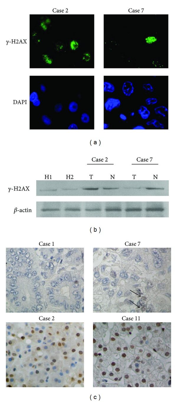
Background: Phosphorylated histone H2AX ( γ -H2AX) is a potential regulator of DNA repair and is a useful tool for detecting DNA damage. To evaluate the clinical usefulness of γ -H2AX in hepatocellular carcinoma (HCC), we measured the level of γ -H2AX in HCC, dysplastic nodule, and nontumorous liver diseases.
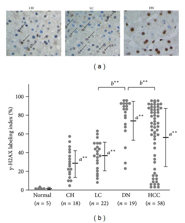
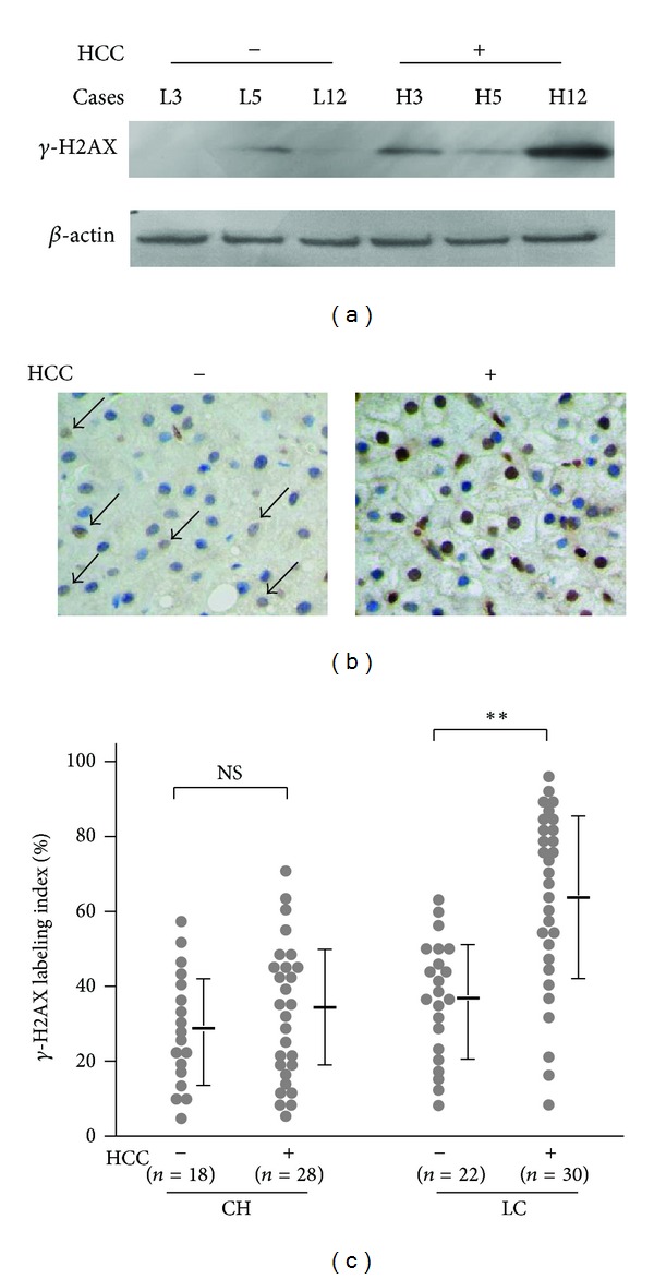
Methods: The level of γ -H2AX was measured by immunohistochemistry in fifty-eight HCC, 18 chronic hepatitis, 22 liver cirrhosis, and 19 dysplastic nodules. Appropriate cases were also examined by fluorescence analysis and western blotting. results: All cases with chronic liver disease showed increased levels of γ -H2AX expression. In 40 (69.9%) of 58 cases with HCC, the labeling index (LI) of γ -H2AX was above 50% and was inversely correlated with the histological grade. Mean γ -H2AX LI was the highest in dysplastic nodule (74.1 ± 22.1%), which was significantly higher than HCC (P < 0.005). Moreover, γ -H2AX was significantly increased in nontumorous tissues of HCC as compared with liver cirrhosis without HCC (62.5 ± 24.7%, from 5.1 to 96.0%, P < 0.005).
Conclusions: γ -H2AX was increased in the preneoplastic lesions of HCC and might be a useful biomarker for predicting the risk of HCC.
Figures
References
-
- Llovet JM, Ricci S, Mazzaferro V, et al. Sorafenib in advanced hepatocellular carcinoma. The New England Journal of Medicine. 2008;359:378–390. - PubMed
-
- Farazi PA, DePinho RA. Hepatocellular carcinoma pathogenesis: from genes to environment. Nature Reviews Cancer. 2006;6(9):674–687. - PubMed
-
- Ince N, Wands JR. The increasing incidence of hepatocellular carcinoma. The New England Journal of Medicine. 1999;340(10):798–799. - PubMed
-
- Matsuda Y, Ichida T, Fukumoto M. Hepatocellular carcinoma and liver transplantation: clinical perspective on molecular targeted strategies. Medical Molecular Morphology. 2011;44:117–124. - PubMed
-
- Della Corte C, Colombo M. Surveillance for hepatocellular carcinoma. Seminars in Oncology. 2012;39:384–398. - PubMed
Publication types
MeSH terms
Substances
LinkOut - more resources
Full Text Sources
Other Literature Sources
