

Clinical and pathological characteristics of organized hematoma
- PMID: 23533421
- PMCID: PMC3606777
- DOI: 10.1155/2013/539642
Clinical and pathological characteristics of organized hematoma
Abstract
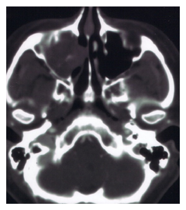
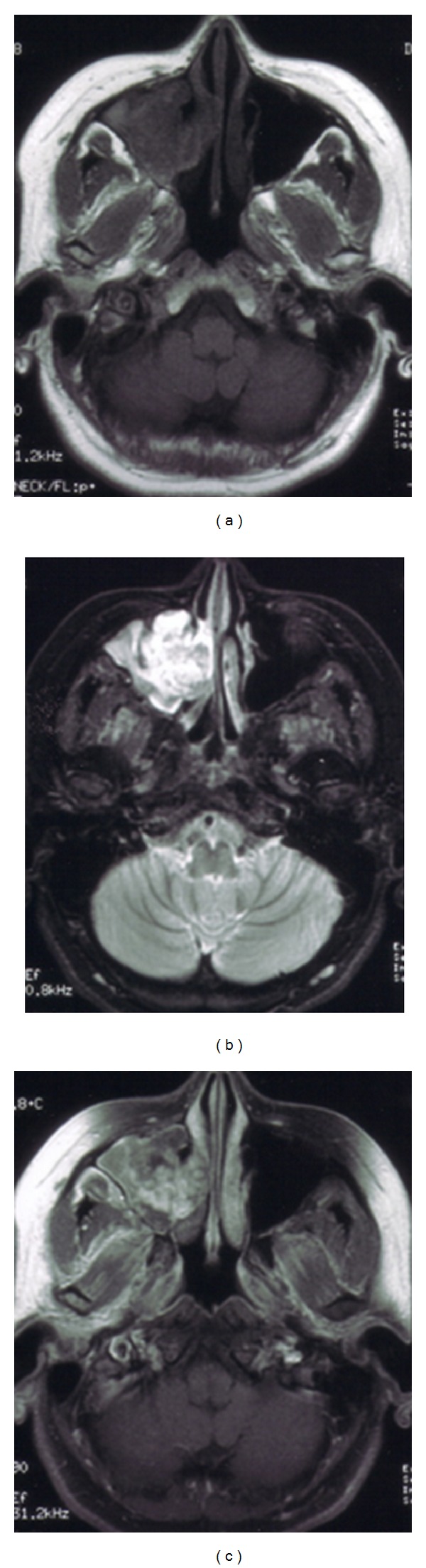
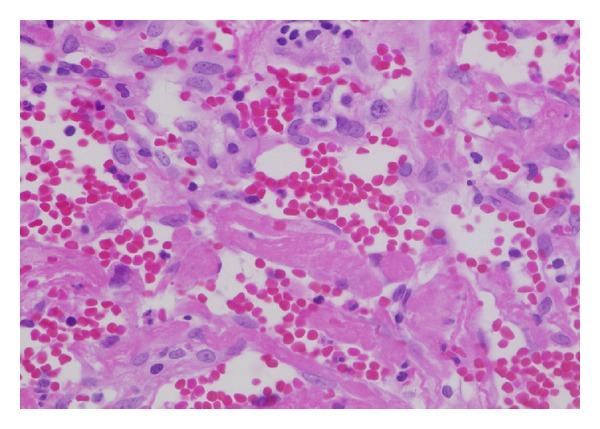
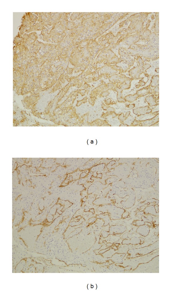
Objective. To study the clinical and pathological characteristics of patients with organized hematoma with malignant features in maxillary sinuses. Subjects and Methods. This was a retrospective study of five patients who were treated surgically for organized hematoma. The preoperative CT and MRI findings were studied clinically. The expressions of CD31, CD34, and periostin in surgical samples were investigated by immunohistochemistry. Results. The clinical features of organized hematoma, such as a mass expanding from the maxillary sinus with bone destruction, resembled those of maxillary carcinoma. However, CT and MRI provided sufficient and useful information to differentiate this condition from malignancy. Surgical resection was the first-line treatment because of the presence of a firm capsule. Characteristic histopathological findings were a mixture of dilated vessels, hemorrhage, fibrin exudation, fibrosis, hyalinization, and neovascularization. The expressions of periostin, CD31, and CD34 were observed in organized hematoma of the maxillary sinus. Conclusion. The expressions of periostin, CD31, and CD34 were observed in organized hematoma of the maxillary sinus. Organized hematoma is characterized pathologically by a mixture of bleeding, dilated vessels, hemorrhage, fibrin exudation, fibrosis, hyalinization, and neovascularization. CT and MRI show heterogeneous findings reflecting a mixture of these pathological entities.
Figures

References
-
- Song HM, Jang YJ, Chung YS, Lee BJ. Organizing hematoma of the maxillary sinus. Otolaryngology—Head and Neck Surgery. 2007;136(4):616–620. - PubMed
-
- Yagisawa M, Ishitoya J, Tsukuda M. Hematoma-like mass of the maxillary sinus. Acta Oto-Laryngologica. 2006;126(3):277–281. - PubMed
-
- Lim M, Lew-Gor S, Beale T, Ramsay A, Lund VJ. Maxillary sinus hematoma. Journal of Laryngology & Otology. 2007;122:210–212. - PubMed
-
- Ricalde RR, Lim ACE, Lopa RAB, Carnate JM. A benign maxillary tumour with malignant features. Rhinology. 2010;48(2):146–149. - PubMed
-
- Lee B-J, Park H-J, Heo S-C. Organized hematoma of the maxillary sinus. Acta Oto-Laryngologica. 2003;123(7):869–872. - PubMed
LinkOut - more resources
Full Text Sources
Other Literature Sources
