

Comparative Evaluation of Human Mesenchymal Stem Cells of Fetal (Wharton's Jelly) and Adult (Adipose Tissue) Origin during Prolonged In Vitro Expansion: Considerations for Cytotherapy
- PMID: 23533440
- PMCID: PMC3603673
- DOI: 10.1155/2013/246134
Comparative Evaluation of Human Mesenchymal Stem Cells of Fetal (Wharton's Jelly) and Adult (Adipose Tissue) Origin during Prolonged In Vitro Expansion: Considerations for Cytotherapy
Abstract
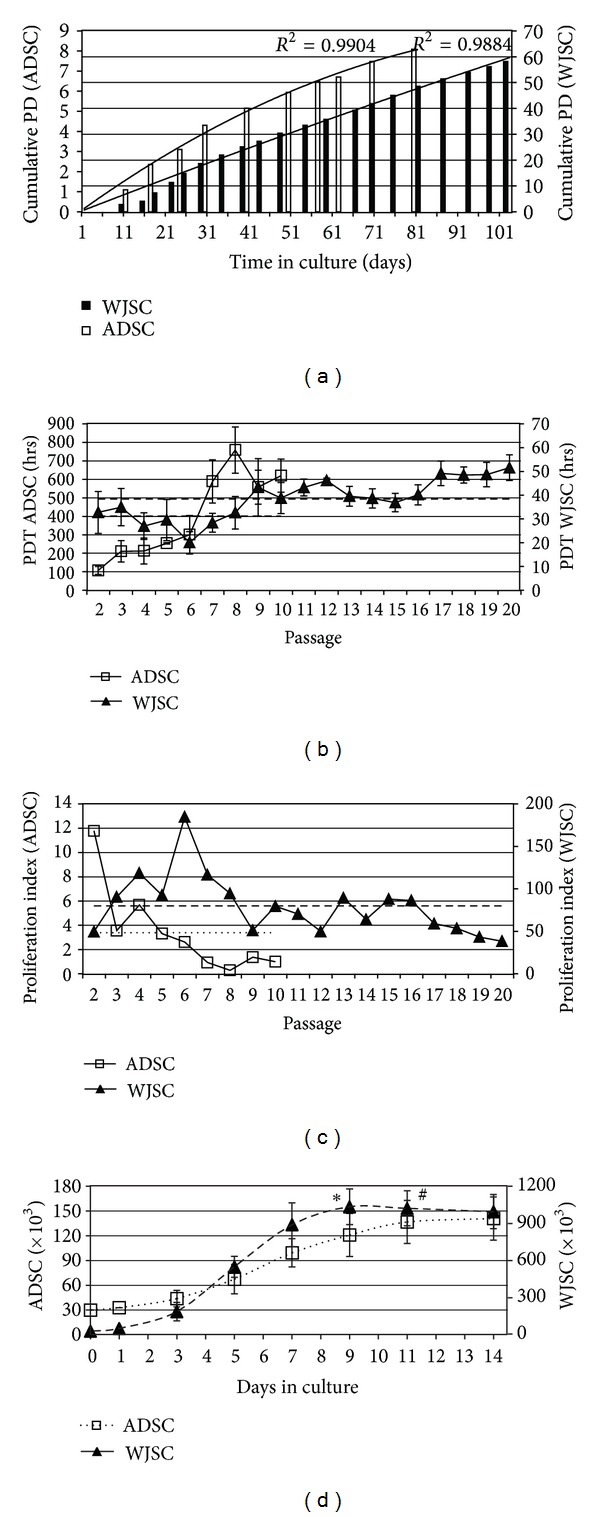
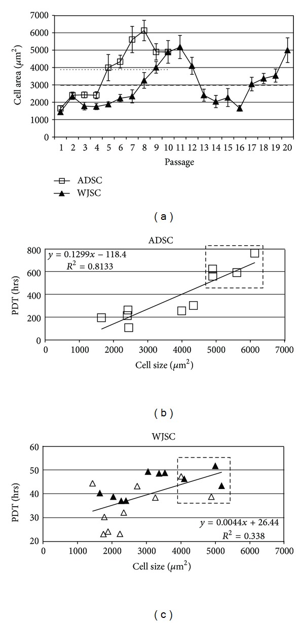
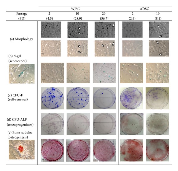
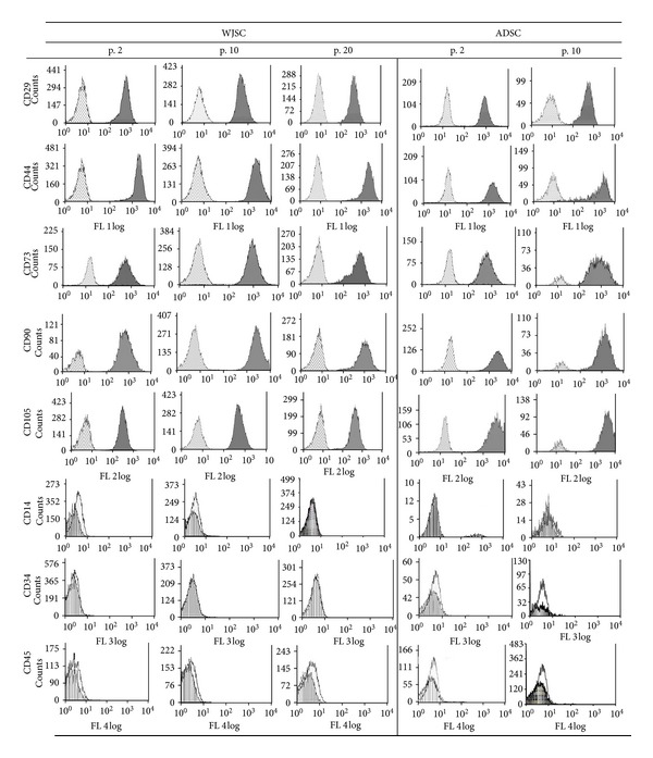
Mesenchymal stem cells (MSCs) are somatic cells with a dual capacity for self-renewal and differentiation, and diverse therapeutic applicability, both experimentally and in the clinic. These cells can be isolated from various human tissues that may differ anatomically or developmentally with relative ease. Heterogeneity due to biological origin or in vitro manipulation is, nevertheless, considerable and may equate to differences in qualitative and quantitative characteristics which can prove crucial for successful therapeutic use. With this in mind, in the present study we have evaluated the proliferation kinetics and phenotypic characteristics of MSCs derived from two abundant sources, that is, fetal umbilical cord matrix (Wharton's jelly) and adult adipose tissue (termed WJSC and ADSC, resp.) during prolonged in vitro expansion, a process necessary for obtaining cell numbers sufficient for clinical application. Our results show that WJSC are derived with relatively high efficiency and bear a substantially increased proliferation capacity whilst largely sustaining the expression of typical immunophenotypic markers, whereas ADSC exhibit a reduced proliferation potential showing typical signs of senescence at an early stage. By combining kinetic with phenotypic data we identify culture thresholds up to which both cell types maintain their stem properties, and we discuss the practical implications of their differences.
Figures
Similar articles
-
Human umbilical cord Wharton's Jelly-derived mesenchymal stem cells differentiation into nerve-like cells.Chin Med J (Engl). 2005 Dec 5;118(23):1987-93. Chin Med J (Engl). 2005. PMID: 16336835
-
Derivation efficiency, cell proliferation, freeze-thaw survival, stem-cell properties and differentiation of human Wharton's jelly stem cells.Reprod Biomed Online. 2010 Sep;21(3):391-401. doi: 10.1016/j.rbmo.2010.04.010. Epub 2010 Apr 24. Reprod Biomed Online. 2010. PMID: 20638335
-
Culturing on Wharton's jelly extract delays mesenchymal stem cell senescence through p53 and p16INK4a/pRb pathways.PLoS One. 2013;8(3):e58314. doi: 10.1371/journal.pone.0058314. Epub 2013 Mar 13. PLoS One. 2013. PMID: 23516461 Free PMC article.
-
Human Wharton's Jelly-Cellular Specificity, Stemness Potency, Animal Models, and Current Application in Human Clinical Trials.J Clin Med. 2020 Apr 12;9(4):1102. doi: 10.3390/jcm9041102. J Clin Med. 2020. PMID: 32290584 Free PMC article. Review.
-
[Research progress of biological characteristics and advantages of Wharton's jelly-mesenchymal stem cells].Zhongguo Xiu Fu Chong Jian Wai Ke Za Zhi. 2011 Jun;25(6):745-9. Zhongguo Xiu Fu Chong Jian Wai Ke Za Zhi. 2011. PMID: 21735792 Review. Chinese.
Cited by
-
Alteration of histone acetylation pattern during long-term serum-free culture conditions of human fetal placental mesenchymal stem cells.PLoS One. 2015 Feb 11;10(2):e0117068. doi: 10.1371/journal.pone.0117068. eCollection 2015. PLoS One. 2015. PMID: 25671548 Free PMC article.
-
Engineered cell-laden thermosensitive poly(N-isopropylacrylamide)-immobilized gelatin microspheres as 3D cell carriers for regenerative medicine.Mater Today Bio. 2022 Apr 19;15:100266. doi: 10.1016/j.mtbio.2022.100266. eCollection 2022 Jun. Mater Today Bio. 2022. PMID: 35517579 Free PMC article.
-
Impact of Antibiotics on the Proliferation and Differentiation of Human Adipose-Derived Mesenchymal Stem Cells.Int J Mol Sci. 2017 Nov 24;18(12):2522. doi: 10.3390/ijms18122522. Int J Mol Sci. 2017. PMID: 29186789 Free PMC article.
-
Antioxidants inhibit cell senescence and preserve stemness of adipose tissue-derived stem cells by reducing ROS generation during long-term in vitro expansion.Stem Cell Res Ther. 2019 Oct 17;10(1):306. doi: 10.1186/s13287-019-1404-9. Stem Cell Res Ther. 2019. PMID: 31623678 Free PMC article.
-
Current View on Osteogenic Differentiation Potential of Mesenchymal Stromal Cells Derived from Placental Tissues.Stem Cell Rev Rep. 2015 Aug;11(4):570-85. doi: 10.1007/s12015-014-9569-1. Stem Cell Rev Rep. 2015. PMID: 25381565 Free PMC article. Review.
References
-
- Nauta AJ, Fibbe WE. Immunomodulatory properties of mesenchymal stromal cells. Blood. 2007;110(10):3499–3506. - PubMed
-
- Patel SA, Sherman L, Munoz J, Rameshwar P. Immunological properties of mesenchymal stem cells and clinical implications. Archivum Immunologiae et Therapiae Experimentalis (Warsz) 2008;56(1):1–8. - PubMed
-
- Phinney DG, Prockop DJ. Concise review: mesenchymal stem/multipotent stromal cells: the state of transdifferentiation and modes of tissue repair—current views. Stem Cells. 2007;25(11):2896–2902. - PubMed
-
- Cavallo C, Cuomo C, Fantini S, et al. Comparison of alternative mesenchymal stem cell sources for cell banking and musculoskeletal advanced therapies. Journal of Cellular Biochemistry. 2011;112(5):1418–1430. - PubMed
LinkOut - more resources
Full Text Sources
Other Literature Sources
