. 2013;8(3):e59374.
doi: 10.1371/journal.pone.0059374. Epub 2013 Mar 22.

The human CD8β M-4 isoform dominant in effector memory T cells has distinct cytoplasmic motifs that confer unique properties

Affiliations

The human CD8β M-4 isoform dominant in effector memory T cells has distinct cytoplasmic motifs that confer unique properties

Deepshi Thakral et al. PLoS One. 2013.

Abstract

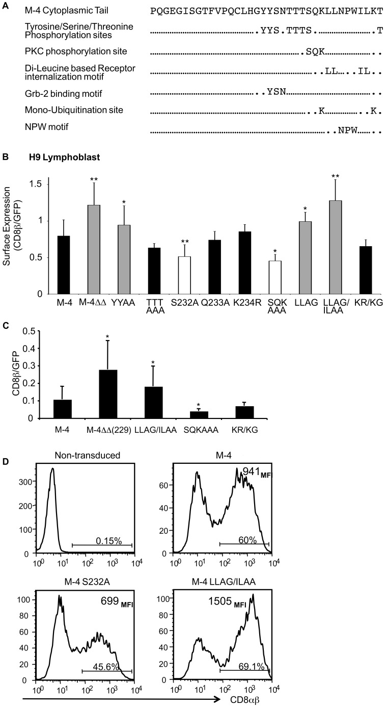
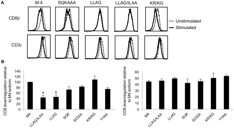
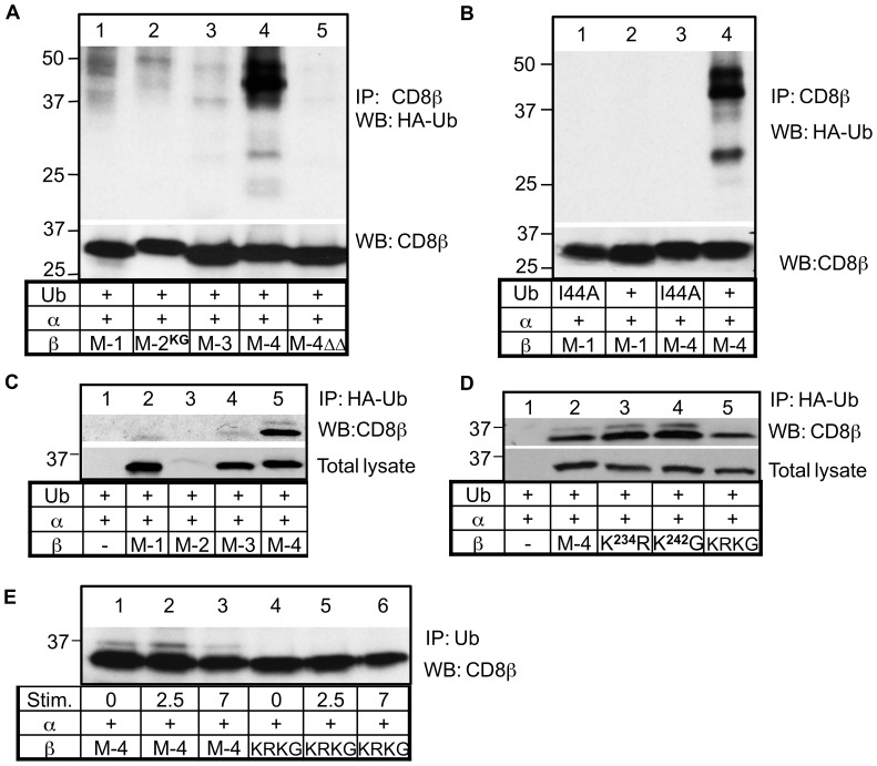
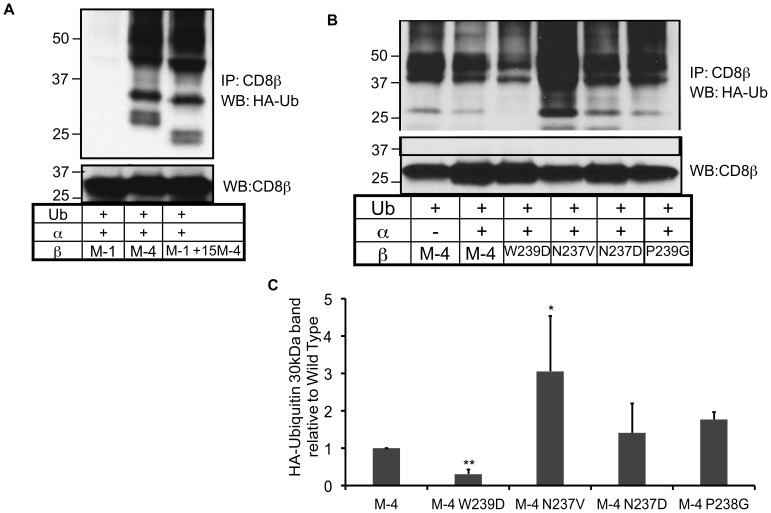
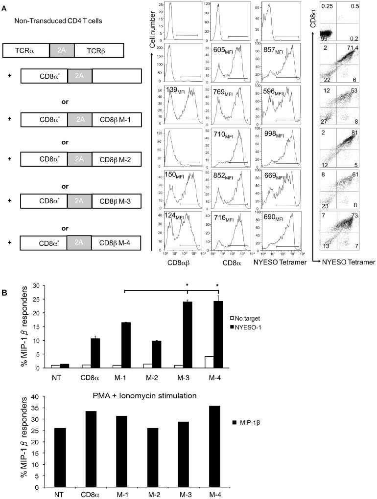
The CD8 co-receptor influences T cell recognition and responses in both anti-tumor and anti-viral immunity. During evolution in the ancestor of humans and chimpanzees, the CD8B gene acquired two additional exons. As a result, in humans, there are four CD8β splice variants (M1 to M4) that differ in their cytoplasmic tails. The M-1 isoform which is the equivalent of murine CD8β, is predominantly expressed in naïve T cells, whereas, the M-4 isoform is predominantly expressed in effector memory T cells. The characteristics of the M-4 isoform conferred by its unique 36 amino acid cytoplasmic tail are not known. In this study, we identified a dihydrophobic leucine-based receptor internalization motif in the cytoplasmic tail of M-4 that regulated its cell surface expression and downregulation after activation. Further the M-4 cytoplasmic tail was able to associate with ubiquitinated targets in 293T cells and mutations in the amino acids NPW, a potential EH domain binding site, either enhanced or inhibited the interaction. In addition, the M-4 tail was itself mono-ubiquitinated on a lysine residue in both 293T cells and a human T cell line. When peripheral blood human T cells expressed CD8αβ M-4, the frequency of MIP-1β secreting cells responding to antigen presenting cells was two-fold higher as compared to CD8αβ M-1 expressing T cells. Thus, the cytoplasmic tail of the CD8β M-4 isoform has unique characteristics, which likely contributed to its selective expression and function in human effector memory T cells.


Conflict of interest statement

Competing Interests: The authors have declared that no competing interests exist.

Figures

Figure 1
Figure 1. Motifs in the cytoplasmic tail of the M-4 isoform that regulate cell surface expression.
(A) Potential amino acid motifs in the cytoplasmic tail of M-4 isoform (www.expasy.org). (B) Quantitative analysis of the surface expression of the M-4 wild-type and mutant proteins expressed in H9 cell line using the anti-CD8β antibody (5F2) by flow cytometry. The amount of CD8β binding was normalized to GFP expression. Each value corresponds to an average of three experiments. The standard deviation and two-population Student’s paired t-test was used to determine statistical differences of the mutants relative to the wild type M-4 isoform, indicated as one star * for p<0.05 and ** p<0.01. (C) Surface expression levels of M-4 wild-type and mutant proteins normalized to GFP expression in primary CD4+ T cells. Peripheral blood CD4+ T cells were stimulated with antibodies against CD3 and CD28 and transduced with lentiviruses expressing GFP and wild type or mutant M-4 proteins. On day 8 cell surface staining with CD8β antibody was analyzed by flow cytometry. The data are the mean +/− S.D. from three independent experiments. Values that are statistically different from the wild type M-4 protein are indicated as * p<0.05 and ** for p<0.01. (D) CD8αβ expression on CD4+ T cells prepared as in (C) expressing M-4 wild-type or mutants S232A or LL235–6AG/IL240–1AA. One representative experiment of five experiments is shown.
Figure 2
Figure 2. Identification of motifs signals in the cytoplasmic tail of M-4 isoform that modulate downregulation from the cell surface.
(A) Receptor downregulation was measured by determining surface expression for CD8β M-4 wild-type and M-4 mutant levels by flow cytometry before and after stimulation of cells with PMA (100 ng/ml) for 60 minutes. One representative of 3 independent experiments is shown. (B) Receptor downregulation relative to CD8β M4 wild type was as in (A) quantitatively represented. Levels of CD3 protein after stimulation are indicated as well. Analyzing three experiments values that are statistically different from the wild type M-4 protein are indicated as * for p<0.05 and ** p<0.01.
Figure 3
Figure 3. Di-leucine motif in the cytoplasmic tail of M-4 isoform regulates cell surface expression.
(A) Histograms showing expression of GFP, CD8αβ heterodimer and total CD8β for M-4 wildtype and mutant proteins analyzed in JM thymoma by flow cytometry. (B) Percentage downregulation of total CD8β in JM expressing M-4 wild type or mutant proteins after stimulation relative to unstimulated cells. (C) Stability studies of CD8β M-4 wild-type and mutants. JM cells treated with cycloheximide, lysed and immunoprecipitated with anti-CD8β antibody were resolved on SDS-PAGE, transferred to a nitrocellulose membrane and probed with anti-CD8β mAb (5F2). A portion of the total cell lysate was analyzed by Western blotting with GAPDH mAb. (D) The CD8β protein level was determined by quantitating the band intensity for each CD8β band and plotting the mount relative to the protein amount at time zero for three independent experiments.
Figure 4
Figure 4. M-4 cytoplasmic tail binds to ubiquitinated proteins and is modified itself by ubiquitination.
(A) HEK-293T cells co-transfected with plasmids expressing HA-tagged ubiquitin, CD8α and each CD8β isoform or mutant proteins. After 48 hours cells were lysed with 1% BRIJ 97 and immunoprecipitated with anti-CD8β mAb, run on a polyacrylamide gel, transferred to a membrane and probed with the anti-HA antibody. Gels were reprobed with an anti-CD8β antibody (5F2). Small differences in size reflect different lengths of the isoforms. (B) Co-transfection of HEK-293T cells with either wild type HA-Ub or I44A mutant of HA-Ub. The immunoprecipitation and Western blotting procedures were performed as in (A). (C) Determination of direct ubiquitination of CD8β isoforms. Cells expressing individual isoforms were immunoprecipitation with anti-HA mAb followed by blotting with anti-CD8β antibody. An aliquot of total cell lysate was similarly analyzed. (D) Effect of lysine mutants on M-4 ubiquitination. Cells expressing M-4 wild-type, single lysine (K234R, K242G) or double lysine (K234R/K242G labeled KRKG) mutants were lysed and protein analyzed as described in (C). (E) The JM T cells expressing CD8α and either CD8β M-4 or the double lysine mutant KRKG were incubated with the anti-CD8α (OKT8) and anti-CD3 (OKT3) mAbs for 0, 2.5 or 7 minutes at 37°C. Cells were lysed, immunoprecipitated with the anti-Ub mAb followed by Western blotting with the anti-CD8β antibody. A representative of three independent experiments is shown in each panel.
Figure 5
Figure 5. The NPW motif in the M-4 cytoplasmic tail mediates binding to ubiquitinated proteins.
(A) HEK-293T cells co-transfected with plasmids expressing HA-tagged ubiquitin, and CD8β wild type M1, M4 or chimeric protein M1 with 15 amino acids of the C terminus of the M4 cytoplasmic tail. After 48 hrs cells were lysed with 1% BRIJ 97, precipitated with anti-CD8β mAb and run on a polyacrylamide gel. Western blotting was performed with the indicated antibody. The membrane was then stripped and re-probed with the anti-CD8β antibody. The experiment was repeated three times. (B) HEK-293T cells co-transfected with plasmids expressing HA-tagged ubiquitin, and CD8β wild type or mutant proteins. The immunoprecipitation and Western blotting experiments were performed as in (A). A representative of four experiments is depicted. (C) The intensity of the 30 kDa band was analyzed and the amount of the 30 kDa band of the wild type relative to each mutant protein is represented. The levels of CD8β protein detected after reprobing with anti-CD8β antibody were used to correct for differences in protein expression between experiments. Student’s paired t-test was used to determine statistical differences of the mutants relative to the wild type M-4 isoform, indicated as one star * for p<0.05.
Figure 6
Figure 6. CD4+ T cells transduced with the M-4 CD8β isoform showed increased frequency of cells producing MIP-1β after stimulation.
Peripheral blood CD4+ T cells were stimulated with anti-CD3 and anti-CD28 antibodies for 24 hours, and then co-transduced with a lentivirus expressing a NY-ESO-1 TCR and another lentivirus expressing CD8α and one of the CD8β isoforms. Cells were stimulated and analyzed for cytokine/chemokine production after day 10–12. (A) Schematic representation of lentiviral vectors used for co-transduction of primary CD4+ T cells is followed by histograms for surface expression of CD8αβ, CD8α and TCR and dot plots showing cell population co-expressing NY-ESO-1 TCR and CD8α. Data were collected by flow cytometry using antibodies against CD8 and an MHC tetramer specific to NY-ESO-1 (NY-ESO- tetramer). Live CD3+ lymphocytes were gated using side vs. forward scatter, anti-CD3 antibody and live/dead cell dye. (B) Frequency of transduced CD4+ T cells producing MIP-1β (top panel) after stimulation with K562 target cells expressing the NY-ESO-1 antigen. T cells without targets served as negative control and cells stimulated with PMA and Ionomycin (bottom panel) were used as positive control. One representative of three independent experiments is shown. Values that are statistically different from the wild type M-1 protein as determined by two-population Student’s paired t-test are indicated as one star (*) for p<0.05.

Similar articles

Cited by

References

    1. Russell JH, Ley TJ (2002) Lymphocyte-mediated cytotoxicity. Annu Rev Immunol 20: 323–370. - PubMed
    1. Yachi PP, Ampudia J, Zal T, Gascoigne NR (2006) Altered peptide ligands induce delayed CD8-T cell receptor interaction–a role for CD8 in distinguishing antigen quality. Immunity 25: 203–211. - PubMed
    1. Holler PD, Kranz DM (2003) Quantitative analysis of the contribution of TCR/pepMHC affinity and CD8 to T cell activation. Immunity 18: 255–264. - PubMed
    1. Purbhoo MA, Boulter JM, Price DA, Vuidepot AL, Hourigan CS, et al. (2001) The human CD8 coreceptor effects cytotoxic T cell activation and antigen sensitivity primarily by mediating complete phosphorylation of the T cell receptor zeta chain. J Biol Chem 276: 32786–32792. - PubMed
    1. Wang H, Kadlecek TA, Au-Yeung BB, Goodfellow HE, Hsu LY, et al. (2010) ZAP-70: an essential kinase in T-cell signaling. Cold Spring Harb Perspect Biol 2: a002279. - PMC - PubMed

Publication types