. 2013:2013:742925.
doi: 10.1155/2013/742925. Epub 2013 Feb 21.

Melanoma-Targeted Chemothermotherapy and In Situ Peptide Immunotherapy through HSP Production by Using Melanogenesis Substrate, NPrCAP, and Magnetite Nanoparticles

Affiliations

Melanoma-Targeted Chemothermotherapy and In Situ Peptide Immunotherapy through HSP Production by Using Melanogenesis Substrate, NPrCAP, and Magnetite Nanoparticles

Kowichi Jimbow et al. J Skin Cancer. 2013.

Abstract

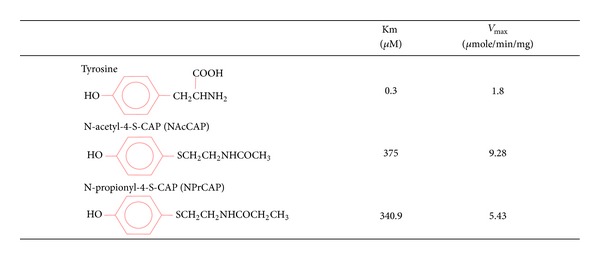
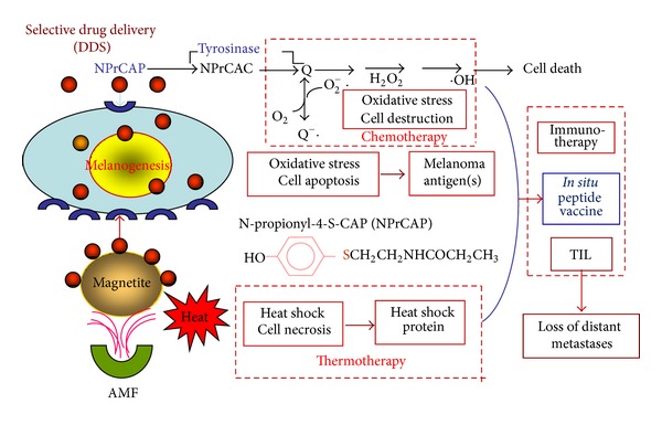
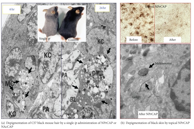
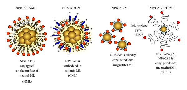
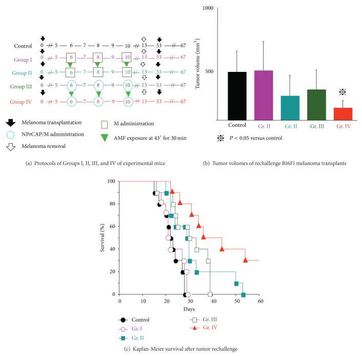
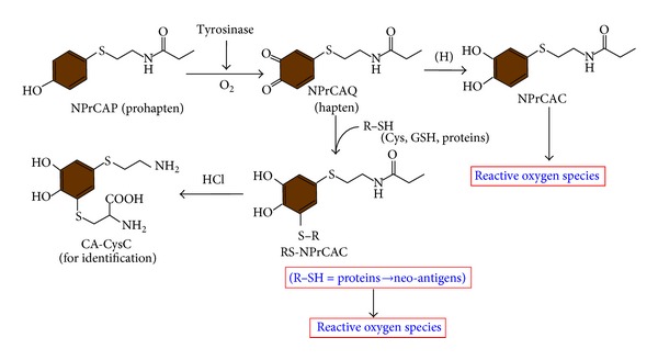
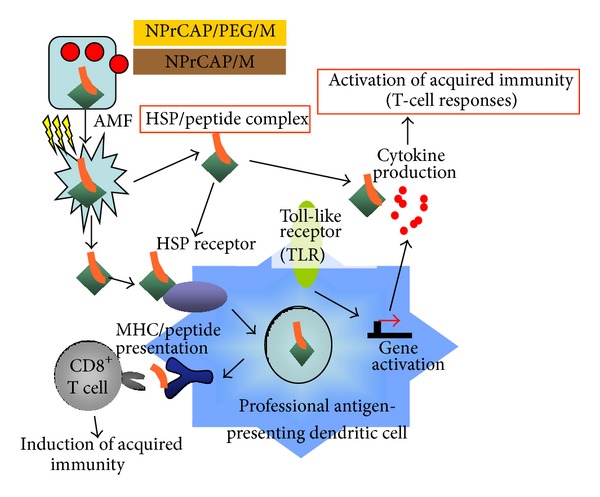
Exploitation of biological properties unique to cancer cells may provide a novel approach to overcome difficult challenges to the treatment of advanced melanoma. In order to develop melanoma-targeted chemothermoimmunotherapy, a melanogenesis substrate, N-propionyl-4-S-cysteaminylphenol (NPrCAP), sulfur-amine analogue of tyrosine, was conjugated with magnetite nanoparticles. NPrCAP was exploited from melanogenesis substrates, which are expected to be selectively incorporated into melanoma cells and produce highly reactive free radicals through reacting with tyrosinase, resulting in chemotherapeutic and immunotherapeutic effects by oxidative stress and apoptotic cell death. Magnetite nanoparticles were conjugated with NPrCAP to introduce thermotherapeutic and immunotherapeutic effects through nonapoptotic cell death and generation of heat shock protein (HSP) upon exposure to alternating magnetic field (AMF). During these therapeutic processes, NPrCAP was also expected to provide melanoma-targeted drug delivery system.


Figures

Figure 1
Figure 1
Synthesis and chemical structures of NAcCAP and NPrCAP and their tyrosinase kinetics.
Figure 2
Figure 2
Strategy for melanogenesis-targeted CTI and in situ peptide vaccine therapy by conjugates of NPrCAP and magnetite nanoparticles with AMF exposure.
Figure 3
Figure 3
Depigmenting effect of NPrCAP. (a) Depigmentation of C57 black mouse hair follicles by a single ip administration of NPrCAP or NAcCAP results in complete loss of melanin pigmentation. Entire coat color changes to silver from black. Electron microscopic observation reveals selective degradation of melanocytes and melanogenic organelles such as early-stage melanosomes at 6 hr after administration. At 24 hr after administration, these melanocytes reveal total degradation. (b) Depigmentation of black skin after topical application of NPrCAP. There is a marked decrease of melanocyte populations after topical application. Electron microscopic observation indicates selective accumulation of NPrCAP in the tyrosinase areas such as in melanosomes and Golgi apparatus as indicated by the deposition of electron dense materials (see arrows).
Figure 4
Figure 4
Conjugates of NPrCAP/magnetite nanoparticles for developing melanogenesis-targeted melanoma nanomedicine.
Figure 5
Figure 5
Melanoma growth and survival of melanoma-bearing mice by CTI therapy using NPrCAP/M with and without AMF exposure. (a) Experimental protocols. (b) Tumor volumes of rechallenge melanoma transplants on day 13 of after transplantation. (c) Kaplan-Meier survival of melanoma-bearing mice after treatment following experimental protocols of Figure 5(a).
Figure 6
Figure 6
Hyperthermia of melanoma cells using B16OVA cells for induction of CTL in CTI therapy. Cytotoxic activity of spleen cells after CTI therapy against B16OVA cells, B16F1 cells, EL4 cells, EL4 cells pulsed with SL8 peptide (OVA-immunodominant peptide), or YAC-1 cells was determined by standard 51Cr-release assay. B16OVA cells were subjected to hyperthermia using NPrCAP/M with AMF exposure in vitro.
Figure 7
Figure 7
NPrCAP-mediated apoptotic cell death of B16F1 melanoma cells. Assay of caspase 3/7 in cells treated with NPrCAP or TRAIL. Cells were cultured in the presence of NPrCAP, TRAIL, or propylene glycol in 96-well plates and then processed for measurement of caspases 3 and 7 using a Caspase-Glo3/7 assay kit. From Ishii-Osai et al. [46].
Figure 8
Figure 8
Tyrosinase activation of NPrCAP (prohapten) and binding of the quinone-hapten NPrCAQ with proteins thorough cysteine residues. Oxidation of NPrCAP with tyrosinase produces the quinone NPrCAQ, which is reduced to the catechol NPrCAC or binds to thiols (cysteine, glutathione, melanosomal proteins). The production of NPrCAQ-thiol adducts can be confirmed by the detection of CA-CysC after acid hydrolysis. NAcCys-NPrCAC is produced by the addition reaction of NAcCys (R-SH) with NPrCAQ. From Ito et al. [47].
Figure 9
Figure 9
Scheme of intracellular hyperthermia using NPrCAP/PEG/M or NPrCAP/M with AMF exposure. NPrCAP/PEG/M nanoparticles are selectively incorporated in melanoma cells. Intracellular hyperthermia can induce necrotic cell death, and adjacent live melanoma cells suffer heat shock, resulting in increased level of intracellular HSP-peptide complexes. Repeated hyperthermia turns heat-shocked cells to necrotic cells, leading to the release of HSP-peptide complexes into extracellular milieu. The released HSPs-peptide complexes are taken up by dendritic cells (DCs). Then, DCs migrate into regional lymph nodes and cross-present HSP chaperoned antigenic peptides to CD8+ T cells in the context of MHC class I molecules, thereby inducing antimelanoma cytotoxic CD8+ T cells.

Similar articles

Cited by

References

    1. de Vries E, van de Poll-Franse LV, Louwman WJ, de Gruijl FR, Coebergh JWW. Predictions of skin cancer incidence in the Netherlands up to 2015. The British Journal of Dermatology. 2005;152(3):481–488. - PubMed
    1. Balch CM, Gershenwald JE, Soong SJ, et al. Final version of 2009 AJCC melanoma staging and classification. Journal of Clinical Oncology. 2009;27(36):6199–6206. - PMC - PubMed
    1. Balch CM, Buzaid AC, Soong SJ, et al. Final version of the American joint committee on cancer staging system for cutaneous melanoma. Journal of Clinical Oncology. 2001;19(16):3635–3648. - PubMed
    1. Reszka K, Jimbow K. Electron donor and acceptor properties of melanin pigments in the skin. In: Fuchs J, Packer L, editors. Oxidative Stress in Dermatology. New York, NY, USA: Marcel Dekker; 1993. pp. 287–320.
    1. Jimbow K, Iwashina T, Alena F, Yamada K, Pankovich J, Umemura T. Exploitation of pigment biosynthesis pathway as a selective chemotherapeutic approach for malignant melanoma. Journal of Investigative Dermatology. 1993;100(2):s231–s238. - PubMed