. 2013:2013:843192.
doi: 10.1155/2013/843192. Epub 2013 Feb 26.

Ovarectomy despite Negative Imaging in Anti-NMDA Receptor Encephalitis: Effective Even Late

Affiliations

Ovarectomy despite Negative Imaging in Anti-NMDA Receptor Encephalitis: Effective Even Late

Anna-Lena Boeck et al. Case Rep Neurol Med. 2013.

Abstract

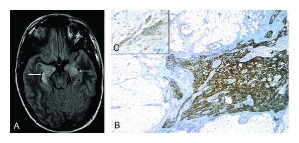
Anti-NMDA receptor (NMDAR) encephalitis is an autoimmune antibody-mediated neuropsychiatric disorder. The disorder is known to be associated with ovarian teratoma and predominantly affects young women. Here, we report the case of a 34-year-old woman with anti-NMDAR encephalitis, in which detailed investigations gave no specific hint for an ovarian teratoma. Despite this, and due to a continuous severe clinical syndrome, an ovarectomy was performed and histological examination revealed an occult teratoma. The ovarectomy led to a remarkable improvement even with a long term intensive care treatment for 11 months. The most important lesson to be learned from this instructive case is that even though none of the investigations was indicative for an ovarian teratoma, including an explorative laparoscopy with biopsy, there still may be an occult ovarian teratoma. This shows that tumour search and diagnosis are extremely important in patients presenting with anti-NMDAR encephalitis, and a laparotomy and ovarectomy is justified. Furthermore, removal of the teratoma even 11 months after a very severe course is still therapeutically effective.


Figures

Figure 1
Figure 1
Axial FLAIR magnetic resonance (MR) imaging (A) shows bilateral hyperintense signal in the mildly swollen medial temporal lobe (arrows). Histological examination (B and C) of the resected ovar revealed a cystic teratoma of 2.9 cm diameter including focal neuronal differentiation of ~0.5 cm diameter. Immunohistochemistry of glial fibrillary acidic protein (B; magnification ×25) and neuron specific enolase (C; magnification ×400) underlined the presence of focal glial and partial neuronal differentiation. Other parts of the teratoma showed squamous epithelial differentiation with hair follicles, sweat as well as sebaceous glands and fat/connective tissue, respiratory epithelia, cartilage as well as osteocytes with bone matrix, and also haematopoiesis.

References

    1. Sansing LH, Tüzün E, Ko MW, Baccon J, Lynch DR, Dalmau J. A patient with encephalitis associated with NMDA receptor antibodies. Nature Clinical Practice Neurology. 2007;3(5):291–296. - PMC - PubMed
    1. Dalmau J, Gleichman AJ, Hughes EG, et al. Anti-NMDA-receptor encephalitis: case series and analysis of the effects of antibodies. The Lancet Neurology. 2008;7(12):1091–1098. - PMC - PubMed
    1. Dalmau J, Lancaster E, Martinez-Hernandez E, Rosenfeld MR, Balice-Gordon R. Clinical experience and laboratory investigations in patients with anti-NMDAR encephalitis. The Lancet Neurology. 2011;10(1):63–74. - PMC - PubMed
    1. Iizuka T, Yoshii S, Kan S, et al. Reversible brain atrophy in anti-NMDA receptor encephalitis: a long-term observational study. Journal of Neurology. 2010;257(10):1686–1691. - PMC - PubMed
    1. Johnson N, Henry C, Fessler AJ, Dalmau J. Anti-NMDA receptor encephalitis causing prolonged nonconvulsive status epilepticus. Neurology. 2010;75(16):1480–1482. - PMC - PubMed

LinkOut - more resources