

"White cord syndrome" of acute tetraplegia after anterior cervical decompression and fusion for chronic spinal cord compression: a case report
- PMID: 23533882
- PMCID: PMC3603640
- DOI: 10.1155/2013/697918
"White cord syndrome" of acute tetraplegia after anterior cervical decompression and fusion for chronic spinal cord compression: a case report
Abstract
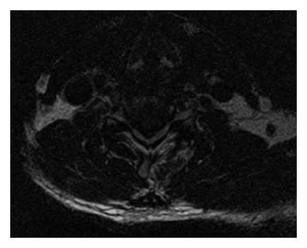
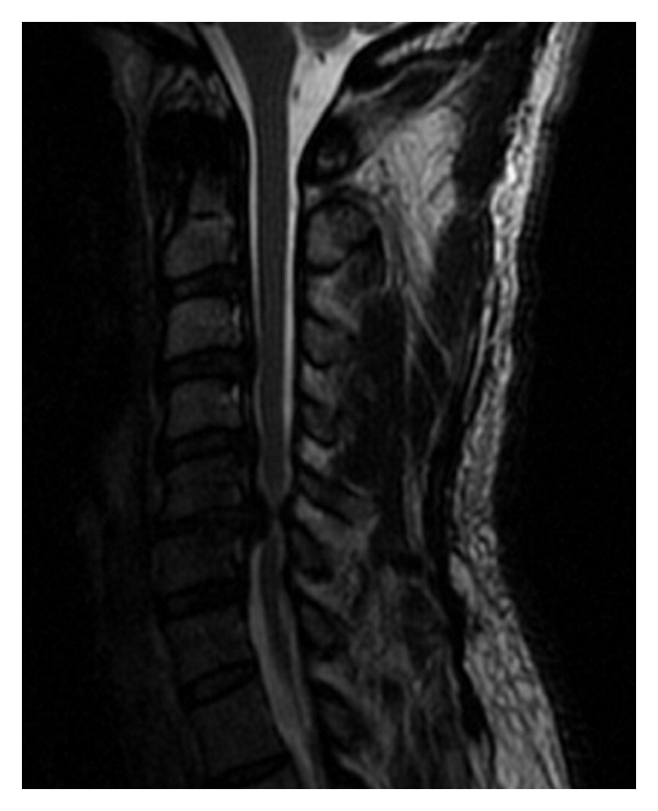
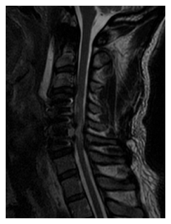
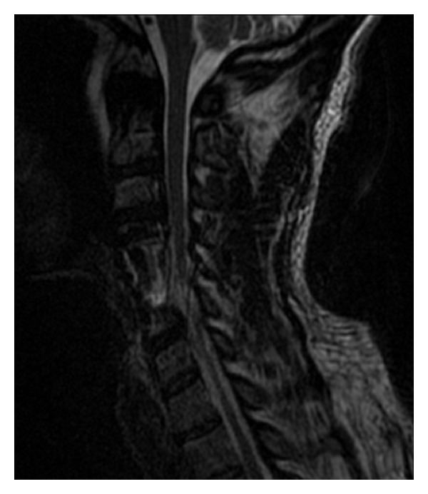
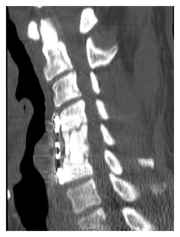
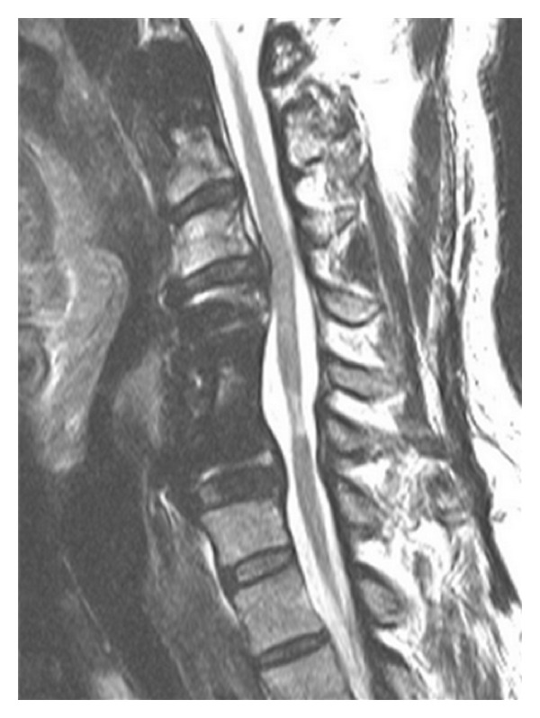
Paralysis is the most feared postoperative complication of ACDF and occurs most often due to an epidural hematoma. In the absence of a clear etiology, inadequate decompression or vascular insult such as ischemia/reperfusion injury are the usual suspects. Herewith we report a case of complete loss of somatosensory evoked potentials (SSEPs) during elective ACDF at C4-5 and C5-6 followed by postoperative C6 incomplete tetraplegia without any discernible technical cause. A postoperative MRI demonstrated a large area of high signal changes on T2-weighted MRI intrinsic to the cord "white cord syndrome" but no residual compression. This was considered consistent with spinal cord gliosis with possible acute edema. The acute decompression of the herniated disc resulted in cord expansion and rush-in reperfusion. We postulate that this may have led to disruption in the blood brain barrier (BBB) and triggered a cascade of reperfusion injuries resulting in acute neurologic dysfunction. At 16 months postoperatively our patient is recovering slowly and is now a Nurick Grade 4.
Figures
References
-
- Mummaneni PV, Kaiser MG, Matz PG, et al. Cervical surgical techniques for the treatment of cervical spondylotic myelopathy. Journal of Neurosurgery. 2009;11(2):130–141. - PubMed
-
- Groff MW, Sriharan S, Lee SM, Maiman DJ. Partial corpectomy for cervical spondylosis. Spine. 2003;28(1):14–19. - PubMed
-
- Jacobs W, Willems PC, van Limbeek J, et al. Single or double-level anterior interbody fusion techniques for cervical degenerative disc disease. Cochrane Database of Systematic Reviews. 2011;1:p. CD004958. - PubMed
-
- Kuntz C, Cramer DE, Maher PC, Pettigrew DB. Major neurologic deficit immediately after adult spinal surgery: incidence and etiology over 10 years at a single training institution. Journal of Spinal Disorders and Techniques. 2009;22(8):565–570. - PubMed
LinkOut - more resources
Full Text Sources
Other Literature Sources
Research Materials
Miscellaneous
