Mixed capillary venous retroperitoneal hemangioma
- PMID: 23533905
- PMCID: PMC3600266
- DOI: 10.1155/2013/258352
Mixed capillary venous retroperitoneal hemangioma
Abstract
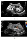
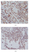
We report a case of mixed capillary venous hemangioma of the retroperitoneum in a 61-year-old man. Abdominal ultrasonography showed a mass to be hypoechoic with increased flow in color Doppler imaging. Dynamic contrast-enhanced computed tomography revealed a centripetal filling-in of the mass, located anterior to the left psoas muscle at the level of sacroiliac joint. On the basis of imaging features, preoperative diagnosis of hemangioma was considered and the mass was excised by laparoscopic method. Immunohistochemical studies were strongly positive for CD31 and CD34, and negative for calretinin, EMA, WT1, HMB45, Ki67, synaptophysin, and lymphatic endothelial cell marker D2-40. Histologically, the neoplasm was diagnosed as mixed capillary venous hemangioma.
Figures


Similar articles
-
Multimodal imaging features of retroperitoneal anastomosing hemangioma: a case report and literature review.Front Oncol. 2023 Oct 25;13:1269631. doi: 10.3389/fonc.2023.1269631. eCollection 2023. Front Oncol. 2023. PMID: 37954079 Free PMC article.
-
Retroperitoneal cavernous hemangioma misdiagnosed as lymphatic cyst: A case report and review of the literature.World J Clin Cases. 2023 May 26;11(15):3560-3570. doi: 10.12998/wjcc.v11.i15.3560. World J Clin Cases. 2023. PMID: 37383918 Free PMC article.
-
Submandibular venous hemangioma: Case report and review of the literature.J Clin Ultrasound. 2015 Oct;43(8):516-9. doi: 10.1002/jcu.22258. Epub 2014 Dec 13. J Clin Ultrasound. 2015. PMID: 25502778 Review.
-
Immunohistochemical investigations of orbital infantile hemangiomas and adult encapsulated cavernous venous lesions (malformation versus hemangioma).Ophthalmic Plast Reconstr Surg. 2013 May-Jun;29(3):183-95. doi: 10.1097/IOP.0b013e31828b0f1f. Ophthalmic Plast Reconstr Surg. 2013. PMID: 23584448
-
Evaluation of thyroid hemangioma by conventional ultrasound combined with contrast-enhanced ultrasound: a case report and review of the literature.J Int Med Res. 2020 Sep;48(9):300060520954718. doi: 10.1177/0300060520954718. J Int Med Res. 2020. PMID: 32972281 Free PMC article. Review.
Cited by
-
A case of retroperitoneal venous malformation resected by laparoscopic surgery.IJU Case Rep. 2022 Jun 1;5(5):369-372. doi: 10.1002/iju5.12491. eCollection 2022 Sep. IJU Case Rep. 2022. PMID: 36090936 Free PMC article.
-
Retroperitoneal infantile hemangioma: a case report and literature review.Discov Oncol. 2024 Aug 27;15(1):373. doi: 10.1007/s12672-024-01260-1. Discov Oncol. 2024. PMID: 39190162 Free PMC article.
-
Retroperitoneal capillary arteriovenous malformation mimicking a malignant neoplasm.IJU Case Rep. 2023 Aug 28;6(6):398-401. doi: 10.1002/iju5.12632. eCollection 2023 Nov. IJU Case Rep. 2023. PMID: 37928304 Free PMC article.
-
Primary anastomosing hemangioma as a preoperative diagnostic mimicker of retroperitoneal cavernous hemangioma: A case report.Oncol Lett. 2024 Apr 9;27(6):254. doi: 10.3892/ol.2024.14386. eCollection 2024 Jun. Oncol Lett. 2024. PMID: 38646490 Free PMC article.
-
Characterization of a gonadal vein Capillary Hemangioma by [68Ga]FAPI-46 and [18 F]FDG PET and immunohistochemistry: a potential pitfall of FAPI PET signal.Eur J Nucl Med Mol Imaging. 2025 Jan;52(2):785-786. doi: 10.1007/s00259-024-06909-1. Epub 2024 Sep 4. Eur J Nucl Med Mol Imaging. 2025. PMID: 39227425 No abstract available.
References
-
- Pérez Martín RN, Estebanez Zarranz J, Velasco Fernández Mdel C, et al. Laparoscopic resection of retroperitoneal venous hemangioma. Journal of Urology. 2004;171, article 336 - PubMed
-
- Igarashi J, Hanazaki K. Retroperitoneal venous hemangioma. American Journal of Gastroenterology. 1998;93(11):2292–2293. - PubMed
-
- Kobayashi H, Kaneko G, Uchida A. Retroperitoneal venous hemangioma. International Journal of Urology. 2010;17(6):585–586. - PubMed
-
- Powis SJ, Rushton DI. A case of retroperitoneal haemangioma. British Journal of Surgery. 1972;59(1):74–76. - PubMed
LinkOut - more resources
Full Text Sources
Other Literature Sources
Research Materials

