. 2013:2013:503725.
doi: 10.1155/2013/503725. Epub 2013 Mar 7.

Infiltration of proinflammatory m1 macrophages into the outer retina precedes damage in a mouse model of age-related macular degeneration

Affiliations

Infiltration of proinflammatory m1 macrophages into the outer retina precedes damage in a mouse model of age-related macular degeneration

Fernando Cruz-Guilloty et al. Int J Inflam. 2013.

Abstract

Age-related macular degeneration (AMD) is a major cause of blindness in the developed world. Oxidative stress and inflammation are implicated in AMD, but precise mechanisms remain poorly defined. Carboxyethylpyrrole (CEP) is an AMD-associated lipid peroxidation product. We previously demonstrated that mice immunized with CEP-modified albumin developed AMD-like degenerative changes in the outer retina. Here, we examined the kinetics of lesion development in immunized mice and the presence of macrophages within the interphotoreceptor matrix (IPM), between the retinal pigment epithelium and photoreceptor outer segments. We observed a significant and time-dependent increase in the number of macrophages in immunized mice relative to young age-matched controls prior to overt pathology. These changes were more pronounced in BALB/c mice than in C57BL/6 mice. Importantly, IPM-infiltrating macrophages were polarized toward the M1 phenotype but only in immunized mice. Moreover, when Ccr2-deficient mice were immunized, macrophages were not present in the IPM and no retinal lesions were observed, suggesting a deleterious role for these cells in our model. This work provides mechanistic evidence linking immune responses against oxidative damage with the presence of proinflammatory macrophages at sites of future AMD and experimentally demonstrates that manipulating immunity may be a target for modulating the development of AMD.


Figures

Figure 1
Figure 1
Immunization with CEP-MSA leads to overt retinal degeneration in aged mice that resembles geographic atrophy with loss of photoreceptor cells, particularly in the BALB/c background. Histology of CEP-MSA versus naïve BALB/c or C57BL/6 eyes at different time points postimmunization. The RPE is located at the lower part of each image. The dark arrows show macrophage-like cells. CEP-MSA BALB/c mice show strong pathology since the early time points, starting with swollen RPE cells and leading to massive geographic atrophy at the late time point, including complete loss of the photoreceptor cells. The kinetics of pathology in CEP-MSA B6 mice is slower, but eventually there are focal lesions of the RPE such as vesiculation, as previously reported [21]. Representative images are shown (from 3–5 mice per group per experiment; two or three independent experiments were performed for each strain). INL: inner nuclear layer; ONL: outer nuclear layer; ROS: rod outer segment; RPE: retinal pigment epithelium. Images were obtained using a 63x oil objective, and the scale marker represents a 20 μm length.
Figure 2
Figure 2
Quantification of retinal pathology and macrophage infiltration in CEP-MSA-immunized mice. (a) Combined pathology score per section (all kinds of lesions and IPM-infiltrating cells). The significant changes due to CEP-MSA immunization occur at the early time point in the BALB/c background, whereas they occur at intermediate time points for B6 mice. (b) CEP-MSA immunization accelerates IPM-infiltrating cell recruitment at early stages of AMD development, prior to major retinal damage. Mean values are shown; error bars represent S.D. (*) denotes statistically significant differences (P < 0.05). Data from one representative experiment was used for this analysis; similar results were obtained in additional repeat experiments. (c) Haematoxylin & Eosin (H&E) staining of frozen sections confirmed the accelerated macrophage recruitment associated with CEP-MSA immunization. 5 sections were scored per mouse, with 3 mice per group at the intermediate time point (d 100–200 p.i.). Mean values are shown; error bars represent S.D.
Figure 3
Figure 3
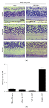
Immune-mediated cellular infiltration and development of outer retinal lesions are specific to genetic background and pigment independent. (a) Histology of CEP-MSA albino B6 mice compared to WT BALB/c and WT B6 at the early time point. Representative images are shown (from 3–5 mice per group per experiment; two or three independent experiments were performed for each strain). INL: inner nuclear layer; ONL: outer nuclear layer; ROS: rod outer segment; RPE: retinal pigment epithelium. Images were obtained using a 63x oil objective, and the scale marker represents a 20 μm length. (b) Quantification shows the lack of pathology at early time points in albino B6 mice, similar to WT B6 and in contrast to WT BALB/c mice. Mean values are shown; error bars represent S.D.
Figure 4
Figure 4
CEP-MSA immunization leads to M1 (TNF-α, IL-12 producing) macrophage recruitment and activation in the subretinal space. (a) Frozen sections followed by immunostaining and confocal microscopy. Surface marker stains were used to identify macrophages as the infiltrate cells. F4/80+ and CD68+ cells are absent from the RPE of naive mice, but they are found in CEP-MSA-immunized BALB/c and B6 mice. (b) Intracellular stains for TNF-α, IL-12, and IL-10 production were used to determine the phenotype of the activated macrophages observed in CEP-MSA BALB/c mice at the intermediate recovery time. IgG2b was used as isotype control. Representative images are shown (from 3–5 mice per group per experiment; two or three independent experiments were performed for each strain). INL: inner nuclear layer; ONL: outer nuclear layer; ROS: rod outer segment; RPE: retinal pigment epithelium; CHO: choroid. The scale marker represents a 20 μm length. (c) Infiltrating macrophages from B6 mice were isolated by laser microdissection, and RNA was obtained for qPCR analysis of gene expression. Relative mRNA (in arbitrary units) was calculated using the 2−ΔCt method with Actin as the calibrator gene. Transcripts for M1 marker genes (IL-1β, TNF, and Ccl2) were detectable in 3 out of 5 CEP-MSA-immunized mice but were not present in CFA age-matched controls (n = 4). M2 marker expression did not correlate with CEP immunization: IL-10 was completely absent, whereas the levels of Arg-1 did not increase. Results are representative of at least two independent laser capture experiments.
Figure 5
Figure 5
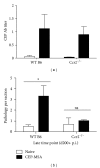
Ccr2 chemokine receptor signaling is required for macrophage recruitment to the outer retina after CEP-MSA immunization. (a) Anti-CEP antibody titers were examined following immunization of WT versus Ccr2−/− B6 mice (n = 5). Naïve mice have no anti-CEP titers. Mean values are shown; error bars represent S.D. (b) Retinal pathology scores for the indicated groups at the late time point (day 200+ p.i.; n = 3). (*) denotes statistically significant difference (P < 0.05); ns: not significant. Data from one representative experiment was used for this analysis; similar results were obtained in a separate independent experiment.

References

    1. Augood CA, Vingerling JR, De Jong PTVM, et al. Prevalence of age-related maculopathy in older Europeans: the European Eye Study (EUREYE) Archives of Ophthalmology. 2006;124(4):529–535. - PubMed
    1. Javitt JC, Zhou Z, Maguire MG, Fine SL, Willke RJ. Incidence of exudative age-related macular degeneration among elderly Americans. Ophthalmology. 2003;110(8):1534–1539. - PubMed
    1. Bressler SB, Maguire MG, Bressler NM, Fine SL. Relationship of drusen and abnormalities of the retinal pigment epithelium to the prognosis of neovascular macular degeneration. Archives of Ophthalmology. 1990;108(10):1442–1447. - PubMed
    1. Sarks SH, Van Driel D, Maxwell L, Killingsworth M. Softening of drusen and subretinal neovascularization. Transactions of the Ophthalmological Societies of the United Kingdom. 1980;100(3):414–422. - PubMed
    1. Holz FG, Bellman C, Staudt S, Schütt F, Völcker HE. Fundus autofluorescence and development of geographic atrophy in age-related macular degeneration. Investigative Ophthalmology and Visual Science. 2001;42(5):1051–1056. - PubMed