Review
. 2013:2013:187204.
doi: 10.1155/2013/187204. Epub 2012 Dec 30.

The inflammatory microenvironment in hepatocellular carcinoma: a pivotal role for tumor-associated macrophages

Affiliations
Review

The inflammatory microenvironment in hepatocellular carcinoma: a pivotal role for tumor-associated macrophages

Daria Capece et al. Biomed Res Int. 2013.

Abstract

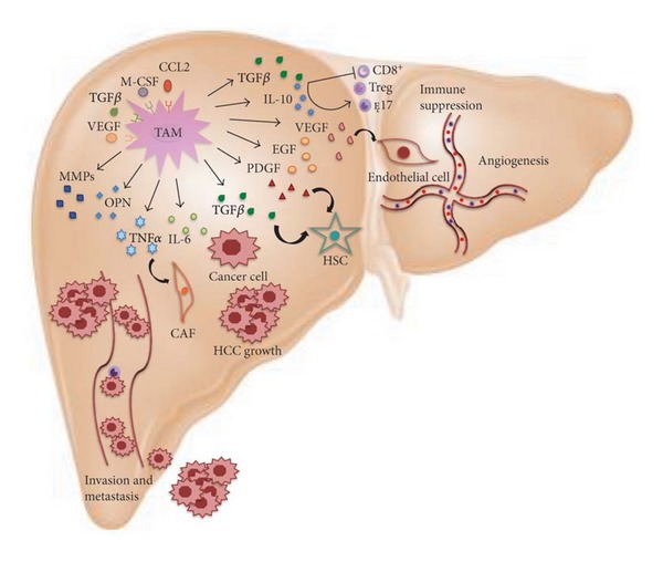
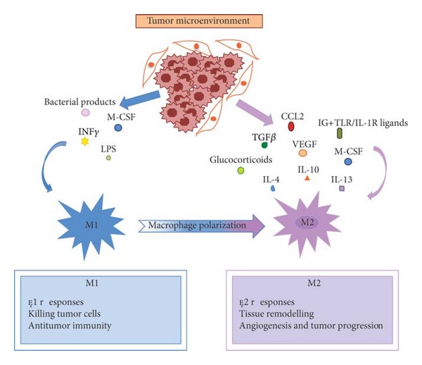
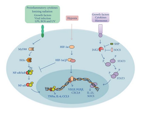
Hepatocellular carcinoma (HCC) is one of the most common and aggressive human cancers worldwide. HCC is an example of inflammation-related cancer and represents a paradigm of the relation occurring between tumor microenvironment and tumor development. Tumor-associated macrophages (TAMs) are a major component of leukocyte infiltrate of tumors and play a pivotal role in tumor progression of inflammation-related cancer, including HCC. Several studies indicate that, in the tumor microenvironment, TAMs acquire an M2-polarized phenotype and promote angiogenesis, metastasis, and suppression of adaptive immunity through the expression of cytokines, chemokines, growth factors, and matrix metalloproteases. Indeed, an established M2 macrophage population has been associated with poor prognosis in HCC. The molecular links that connect cancer cells and TAMs are not completely known, but recent studies have demonstrated that NF-κB, STAT-3, and HIF-1 signaling pathways play key roles in this crosstalk. In this paper, we discuss the current knowledge about the role of TAMs in HCC development, highlighting the role of TAM-derived cytokines, chemokines, and growth factors in the initiation and progression of liver cancer and outlining the signaling pathways involved in the interplay between cancer cells and TAMs.


Figures

Figure 1
Figure 1
The roles of tumor-associated macrophages (TAMs) in liver cancer. TAMs promote hepatocellular carcinoma (HCC) growth, angiogenesis, invasion, and metastasis, as well as the suppression of antitumor immune response by interacting with both stromal and cancer cells within the tumor microenvironment. TAMs are recruited in HCC milieu by M-CSF, CCL2, VEGF, and TGFβ, and they, in turn, release many cytokines, chemokines and growth factors, which are implicated in such crosstalk. In particular, IL-6 and TGFβ favor tumor growth, whereas TNFα, OPN, MMPs, and IL-6 are involved in invasion and metastasis; TGFβ, in concert with IL-10, promotes the suppression of antitumor immune response. Finally, angiogenesis is induced by several molecules, including VEGF, EGF, PDGF, and TGFβ. Refer to the text for abbreviations.
Figure 2
Figure 2
The phenotypic polarization of macrophages in the tumor microenvironment. Macrophages can be schematically classified into two main classes depending on their phenotypic polarization: macrophages mount M1 phenotype in response to M-CSF, INFγ, LPS and other microbial products, whereas they differentiate into M2 in the presence of TGFβ, VEGF, CCL2, M-CSF, IL-4, IL-10, IL-13, glucocorticoids and immune complexes/TLR ligands. M1 and M2 display different functions. M1 macrophages are able to trigger Th1 immune response and exert cytotoxic activity towards ingested microorganisms and cancer cells. M2 macrophages activate Th2 immune response and promote angiogenesis, tissue remodeling, and tumor progression. Refer to the text for abbreviations.
Figure 3
Figure 3
Schematic representation of NF-κB, HIF-1, and STAT-3 signaling pathways linking inflammation and liver cancer. Proinflammatory signals bind to their corresponding receptors, leading to the recruitment of receptor-associated proteins, such as MyD-88. In turn, these associated proteins trigger a phosphorylation cascade that leads to activation of the IKK complex, which is responsible for the phosphorylation of the IκB protein. Phosphorylated IκB is degraded by proteasome, thereby allowing free NF-κB dimers to translocate to the nucleus and transactivate target genes, such as TNFα, IL-6, and CCL3. HIF-1α is the central regulator of the hypoxic response. HIF-1α is activated by hypoxia and its activity progressively increases with a decrease in O2 gradient. Heterodimerization of HIF-1α with HIF-1β allows for DNA binding to hypoxia response elements (HRE) and transactivation of its target genes, such as VEGF, FGFβ, and CXCL8. Cytokine, hormone and growth factor stimulation activates JAK2, which in turn phosphorylates STAT3, allowing its dimerization, nuclear translocation, and transactivation of target genes, such as IL-23. STAT-3 signaling is turned off by protein inhibitors, such as SHP and SOCS, which are induced by STAT-3 in a negative feedback loop. Refer to the text for abbreviations.

References

    1. Yang JD, Nakamura I, Roberts LR. The tumor microenvironment in hepatocellular carcinoma: current status and therapeutic targets. Seminars in Cancer Biology. 2011;21(1):35–43. - PMC - PubMed
    1. Bruix J, Boix L, Sala M, Llovet JM. Focus on hepatocellular carcinoma. Cancer Cell. 2004;5(3):215–219. - PubMed
    1. Berasain C, Castillo J, Perugorria MJ, Latasa MU, Prieto J, Avila MA. Inflammation and liver cancer: new molecular links. Annals of the New York Academy of Sciences. 2009;1155:206–221. - PubMed
    1. Porta C, Riboldi E, Sica A. Mechanisms linking pathogens-associated inflammation and cancer. Cancer Letters. 2011;305(2):250–262. - PubMed
    1. Budhu A, Xin WW. The role of cytokines in hepatocellular carcinoma. Journal of Leukocyte Biology. 2006;80(6):1197–1213. - PubMed

Publication types

MeSH terms

LinkOut - more resources