Is magnetic resonance imaging sufficient to diagnose rudimentary uterine horn? A case report and review of the literature
- PMID: 23535077
- PMCID: PMC3720688
- DOI: 10.1016/j.jmig.2013.01.012
Is magnetic resonance imaging sufficient to diagnose rudimentary uterine horn? A case report and review of the literature
Abstract
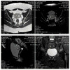
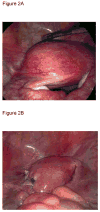
Imaging is often part of the evaluation of gynecologic disorders, with transvaginal ultrasound being the most frequently used imaging modality. Although laparoscopy, hysterosalpingography, and hysteroscopy can add diagnostic accuracy, they are invasive and costly. Magnetic resonance imaging (MRI) has been increasingly used because it is both noninvasive and highly accurate. Although MRI is more expensive than ultrasound, it is less so than surgery. Given the demonstrated accuracy of MRI in assessing müllerian anomalies, additional imaging is not often sought once an MRI diagnosis is made. However, when imaging findings are not pathognomonic via MRI or otherwise, inaccurate diagnoses and their consequences may occur. We describe the case of a 21-year-old woman with unilateral dysmenorrhea whose MRI features suggested a unicornuate uterus with a hematometrous noncommunicating horn although laparoscopy ultimately revealed a necrotic myoma without an accompanying müllerian anomaly.
Keywords: Laparoscopy; Leiomyoma; Magnetic resonance imaging; Myomectomy; Müllerian anomaly; Unicornuate uterus.
Published by Elsevier Inc.
Figures


References
-
- Raga F, Bauset C, Remohi J, et al. Reproductive impact of congenital Mullerian Anomalies. Hum Reprod. 1997;12:2277–81. - PubMed
-
- The American Fertility Society classifications of adnexal adhesion, distal tubal occlusion, tubal occlusion secondary to tubal ligation, tubal pregnancies, Mullerian anomalies and intrauterine adhesions. Fertil Steril. 1988;49:944. - PubMed
-
- Agarwal M, Das A, Singh AS. Dysmenorrhea due to a rare Mullerian anomaly. Niger J Clin Pract. 2011;14:377–9. - PubMed
-
- Strawbridge LC, Crouch NS, Cutner AS, et al. Obstructive Mullerian Anomalies and Modern Laparoscopic Management. J Pediatr Adolesc Gynecol. 2007;20:195–200. - PubMed
-
- Pellerito JS, McCarthy SM, Dovie MB, Glickman MG, DeCherney AH. Diagnosis of uterine anomalies: relative accuracy of MR imaging, endovaginal sonography, and hysterosalpingography. Radiology. 1992;183:795–800. - PubMed
Publication types
MeSH terms
Grants and funding
LinkOut - more resources
Full Text Sources
Other Literature Sources
Medical

