Review
. 2013 Jul;1834(7):1271-8.
doi: 10.1016/j.bbapap.2013.03.007. Epub 2013 Mar 25.

PKA: lessons learned after twenty years

Affiliations
Review

PKA: lessons learned after twenty years

Susan S Taylor et al. Biochim Biophys Acta. 2013 Jul.

Abstract

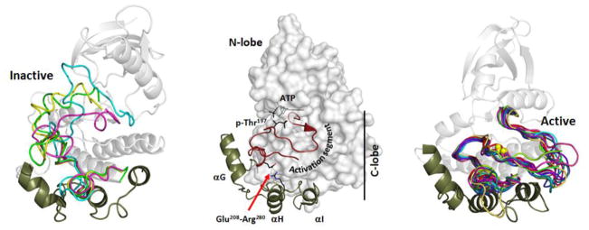
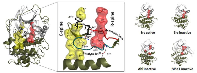
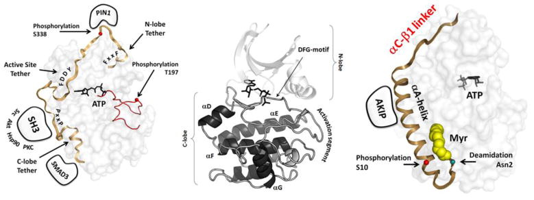
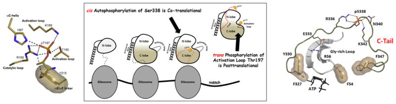
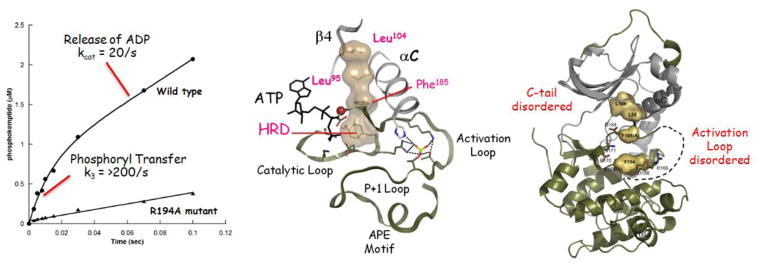
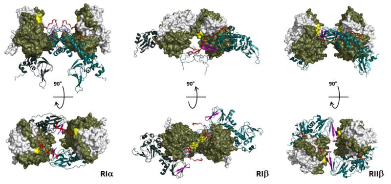
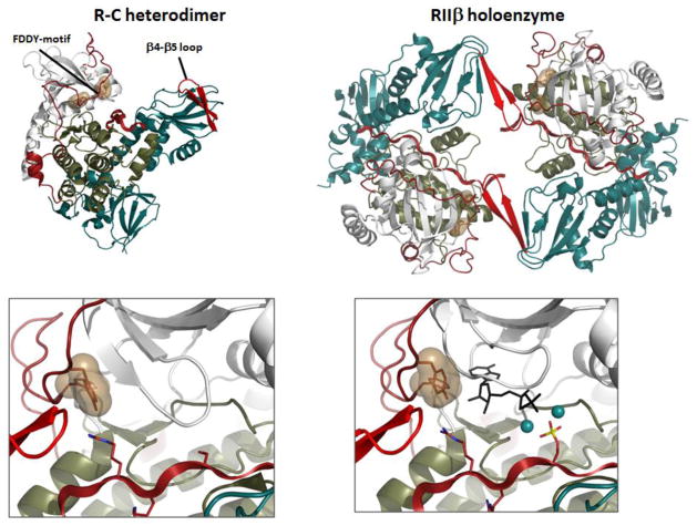
The first protein kinase structure, solved in 1991, revealed the fold that is shared by all members of the eukaryotic protein kinase superfamily and showed how the conserved sequence motifs cluster mostly around the active site. This structure of the PKA catalytic (C) subunit showed also how a single phosphate integrated the entire molecule. Since then the EPKs have become a major drug target, second only to the G-protein coupled receptors. Although PKA provided a mechanistic understanding of catalysis that continues to serve as a prototype for the family, by comparing many active and inactive kinases we subsequently discovered a hydrophobic spine architecture that is a characteristic feature of all active kinases. The ways in which the regulatory spine is dynamically assembled is the defining feature of each protein kinase. Protein kinases have thus evolved to be molecular switches, like the G-proteins, and unlike metabolic enzymes which have evolved to be efficient catalysis. PKA also shows how the dynamic tails surround the core and serve as essential regulatory elements. The phosphorylation sites in PKA, introduced both co- and post-translationally, are very stable. The resulting C-subunit is then packaged as an inhibited holoenzyme with cAMP-binding regulatory (R) subunits so that PKA activity is regulated exclusively by cAMP, not by the dynamic turnover of an activation loop phosphate. We could not understand activation and inhibition without seeing structures of R:C complexes; however, to appreciate the structural uniqueness of each R2:C2 holoenzyme required solving structures of tetrameric holoenzymes. It is these tetrameric holoenzymes that are localized to discrete sites in the cell, typically by A Kinase Anchoring Proteins where they create discrete foci for PKA signaling. Understanding these dynamic macromolecular complexes is the challenge that we now face. This article is part of a Special Issue entitled: Inhibitors of Protein Kinases (2012).


Figures

Fig 1
Fig 1. Structural features that distinguish the Eukaryotic Protein Kinases (EPKs) from the Eukaryote-Like Protein Kinases (ELKs)
The N and C-lobe architecture is maintained in both the ELKs and the EPKs; however, there are two features that are unique to the EPKs. There is a large and highly regulated activation segment (shown in red) that is inserted between β strand 9 and the α F-Helix in the C-Lobe. The conformation of this segment is typically regulated by phosphorylation. The activation loop phosphorylation site for PKA, Thr197, is indicated. The second structural feature that is unique to the EPKs is a helical bundle that includes the αG, αH and αI Helices. This serves most often as a docking site for protein substrates and is thought to be coupled allosterically to the active site [43]. The helix bundle is directly linked to the activation segment by a conserved ion pair between Arg280 between the αH and αI helices and Glu208 in the activation segment.
Fig. 2
Fig. 2. Architecture of the kinase core is defined by two hydrophobic spines
The two lobes of the protein kinase core are linked by two hydrophobic spines, a catalytic spine (C-spine; yellow) there the spine is completed by the adenine ring of ATP and a regulatory spine (R-spine; red() that is assembled in a dynamic manner, typically by phosphorylation of the activation loop. The two spines are intact in every active kinase whereas in inactive kinases the R-spine is broken. Examples showing how the R-spine is assembled in an active kinase (Src:pdb1y57) and broken in an inactive kinase are shown on the right for Src (PDB2src), Abl (PDB1Opj) and Msk (PDB1vzO).
Fig. 3
Fig. 3. N-terminal and C-terminal tails flank the kinase core of PKA
The conserved N and C-lobes of the PKA kinase core are shown in the center. This core is flanked by dynamic tails that serve as both cis and trans regulatory elements. On the right is the N-terminal tail which is myristylated at the N-terminal Glycine. On the left is the C-tail which is a conserved feature of all AGC kinases.
Fig. 4
Fig. 4. Assembly of an active PKA catalytic subunit
The PKA catalytic subunit is assembled as an active kinase by the phosphorylation of two residues, Thr197 in the activation loop and Ser338 in the C-terminal tail. The two phosphates serve essential but different roles in the mature enzyme. The activation loop phosphate (left) interacts with many residues and is essential for the fully active enzyme and for stability. Ser338 is part of the loop that wraps around the N-lobe and controls the C-Helix. The C-tail is anchored to the N-Lobe by a hydrophobic motif at the C-terminus (Phe-X-X-Phe) that binds to the C-Helix in the N-lobe while another C-Tail motif (FDDY) is anchored to the ATP. Both motifs as well as a phosphorylation site between the two sites is a conserved feature of all AGC kinases. Ser338 is phosphorylated co-translationally while the protein is still on the ribosome while the phosphate is added to Thr 197 by a trans-phosphorylation event after the protein leaves the ribosome.
Fig. 5
Fig. 5. Effects of activation loop phosphorylation on the activity and structure of the PKA catalytic subunit
Phosphorylation of the activation loop assembles the R-spine (center) while removal of the activation loop phosphate eliminates the pre-steady state burst o catalytic activity (left) and leads to major disorder in the structure. Specifically the activation loop becomes disordered and the portion of the C-Tail that binds to ATP and is referred to as the active site tether is also disordered.
Fig. 6
Fig. 6. Tetrameric holoenzyme structures of RIa, RIb and RIIb
The structures all reveal a twofold symmetry that was not seen in earlier structures of R:C heterodimers; however, the symmetry is different for each holoenzyme. Although the domain organization of each R-subunit is conserved, the quaternary structures are quite different.
Figure 7
Figure 7. Structure of the RIIb holoenzyme traps both products in the crystal lattice
The tetrameric RIIb holoenzyme is assembled as two R:C heterodimers . the dimer interface in the holoenzyme is created by the b4-b5 loop in the CNB-A domain of the R-subunit and the FDDY motif in the C-terminal tail of the C-subunit. The C-Tail is in a fully closed conformation in the tetramer even though no nucleotide is present. When MgATP was added to the crystals, the phosphate was transferred and both products, ADP and the phosphorylated RIIb subunit including two Mg2+ ions were trapped in the crystal lattice.

References

    1. Taylor SS, Keshwani MM, Steichen JM, Kornev AP. Evolution of the eukaryotic protein kinases as dynamic molecular switches. Philos T R Soc B. 2012;367:2517–2528. - PMC - PubMed
    1. Masterson LR, Cembran A, Shi L, Veglia G. Allostery and binding cooperativity of the catalytic subunit of protein kinase A by NMR spectroscopy and molecular dynamics simulations. Adv Protein Chem Struct Biol. 2012;87:363–389. - PMC - PubMed
    1. Masterson LR, Cheng C, Yu T, Tonelli M, Kornev A, Taylor SS, Veglia G. Dynamics connect substrate recognition to catalysis in protein kinase A. Nat Chem Biol. 2010;6:821–828. - PMC - PubMed
    1. Masterson LR, Shi L, Metcalfe E, Gao J, Taylor SS, Veglia G. Dynamically committed, uncommitted, and quenched states encoded in protein kinase A revealed by NMR spectroscopy. Proc Natl Acad Sci U S A. 2011 - PMC - PubMed
    1. Krebs EG, Graves DJ, Fischer EH. Factors affecting the activity of muscle phosphorylase B kinase. Journal of Biological Chemistry. 1959;234:2867–2873. - PubMed

Publication types

LinkOut - more resources