

Terminals of the major thalamic input to visual cortex are devoid of synapsin proteins
- PMID: 23535254
- PMCID: PMC3953556
- DOI: 10.1016/j.neuroscience.2013.03.031
Terminals of the major thalamic input to visual cortex are devoid of synapsin proteins
Abstract
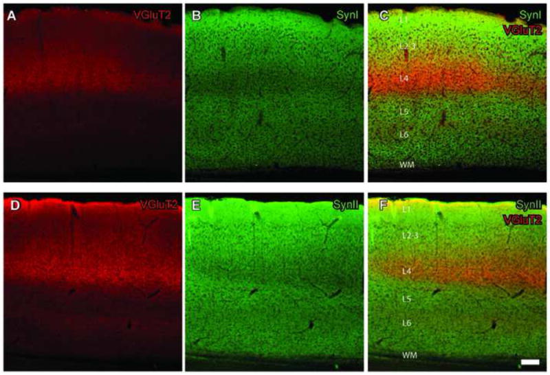
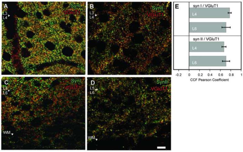
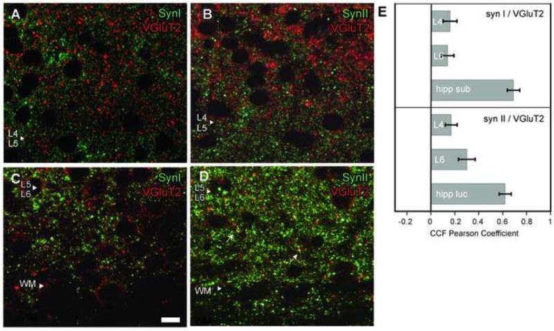
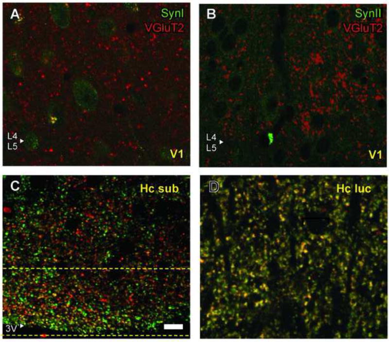
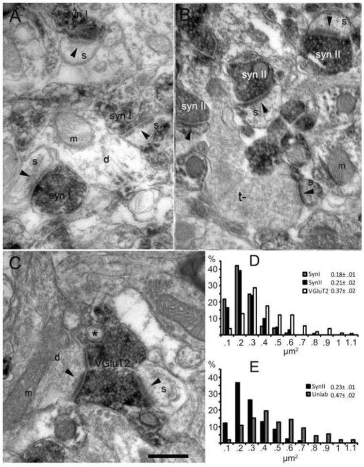
Synapsins are nerve-terminal proteins that are linked to synaptic transmission and key factors in several forms of synaptic plasticity. While synapsins are generally assumed to be ubiquitous in synaptic terminals, whether they are excluded from certain types of terminals is of interest. In the visual pathway, synapsins are lacking in photoreceptor and bipolar cell terminals as well as in retinogeniculate synapses. These are the terminals of the first three feedforward synapses in the visual pathway, implying that lack of synapsins may be a common property of terminals that provide the primary driver activity onto their postsynaptic neurons. To further investigate this idea, we studied the fourth driver synapse, thalamocortical synapses in visual cortex, using glutamatergic terminal antibody markers anti-VGluT1 and VGluT2, anti-Synapsin I and II, and confocal microscopy to analyze co-localization of these proteins in terminals. We also used pre-embedding immunocytochemical labeling followed by electron microscopy to investigate morphological similarities or differences between terminals containing synapsins or VGluT2. In visual cortex, synapsin coincided extensively with non-TC-neuron marker, VGluT1, while thalamocortical terminal marker VGluT2 and synapsin overlap was sparse. Morphologically, synapsin-stained terminals were smaller than non-stained, while VGluT2-positive thalamocortical terminals constituted the largest terminals in cortex. The size discrepancy between synapsin- and VGluT2-positive terminals, together with the complementary staining patterns, indicates that thalamocortical synapses are devoid of synapsins, and support the hypothesis that afferent sensory information is consistently transmitted without the involvement of synapsins. Furthermore, VGluT2 and synapsins were colocalized in other brain structures, suggesting that lack of synapsins is not a property of VGluT2-containing terminals, but a property of primary driver terminals in the visual system.
Copyright © 2013 IBRO. Published by Elsevier Ltd. All rights reserved.
Figures
References
-
- Ahmed B, Anderson JC, Douglas RJ, Martin KA, Nelson JC. Polyneuronal innervation of spiny stellate neurons in cat visual cortex. J Comp Neurol. 1994;341:39–49. - PubMed
-
- Ahmed B, Anderson JC, Martin KA, Nelson JC. Map of the synapses onto layer 4 basket cells of the primary visual cortex of the cat. J Comp Neurol. 1997;380:230–242. - PubMed
-
- Barroso-Chinea P, Castle M, Aymerich MS, Pérez-Manso M, Erro E, Tuñon T, Lanciego JL. Expression of the mRNAs encoding for the vesicular glutamate transporters 1 and 2 in the rat thalamus. J Comp Neurol. 2007;501:703–715. - PubMed
-
- Bolte S, Cordelieres FP. A guided tour into subcellular colocalization analysis in light microscopy. J Microsc. 2006;224:213–32. - PubMed
Publication types
MeSH terms
Substances
Grants and funding
LinkOut - more resources
Full Text Sources
Other Literature Sources
Molecular Biology Databases
