. 2013:4:1626.
doi: 10.1038/ncomms2623.

Sulfhydration mediates neuroprotective actions of parkin

Affiliations

Sulfhydration mediates neuroprotective actions of parkin

M Scott Vandiver et al. Nat Commun. 2013.

Abstract

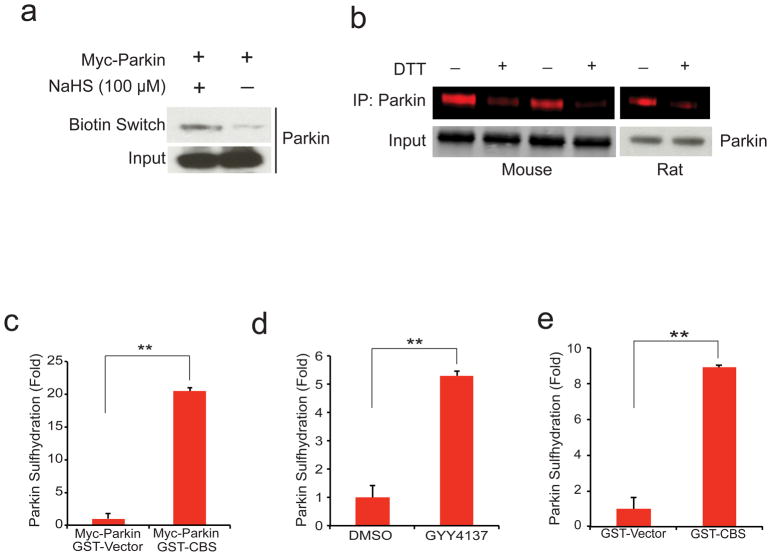
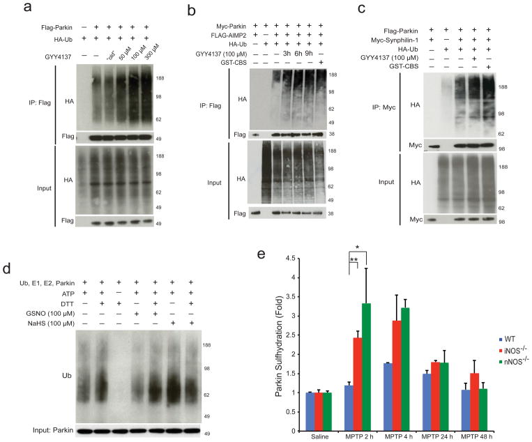
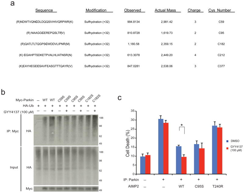
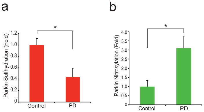
Increases in S-nitrosylation and inactivation of the neuroprotective ubiquitin E3 ligase, parkin, in the brains of patients with Parkinson's disease are thought to be pathogenic and suggest a possible mechanism linking parkin to sporadic Parkinson's disease. Here we demonstrate that physiologic modification of parkin by hydrogen sulfide, termed sulfhydration, enhances its catalytic activity. Sulfhydration sites are identified by mass spectrometry analysis and are investigated by site-directed mutagenesis. Parkin sulfhydration is markedly depleted in the brains of patients with Parkinson's disease, suggesting that this loss may be pathologic. This implies that hydrogen sulfide donors may be therapeutic.


Conflict of interest statement

Competing Financial Interests:

The authors declare no competing financial interests.

Figures

Figure 1
Figure 1. Parkin is physiologically sulfhydrated
(a) Parkin expressed in HEK293 cells is sulfhydrated by the H2S donor NaHS as detected by the modified biotin switch method. (b) Endogenous parkin is basally sulfhydrated in both mouse brain and rat striatum as detected by the maleimide assay in which loss of red fluorescence signal following DTT treatment indicates sulfhydration of the protein. (c) Sulfhydration of myc-parkin overexpressed in HEK293 cells is enhanced almost 20 fold upon overexpression of GST-CBS, one of the principle H2S producers in the brain. n=3, P<0.01 via one-way ANOVA. (d) Endogenous parkin sulfhydration in SH-SY5Y cells is enhanced over 5 fold upon treatment with 100 μM GYY4137, a hydrogen sulfide donor. n=3, P<0.01 via one-way ANOVA. (e) Endogenous parkin sulfhydration in SH-SY5Y cells is enhanced over 8 fold by overexpression of GST-CBS. n=3, P<0.01 via one-way ANOVA. All data expressed as mean ± s.e.m.
Figure 2
Figure 2. Hydrogen sulfide enhances parkin E3 ligase activity via sulfhydration
(a) Parkin E3 ligase activity in HEK293 cells is augmented by GYY4137 in a dose dependent manner, but not by “old” GYY4137, which was exposed to air overnight in PBS, and is subsequently unable to donate H2S. (b) Parkin activity, measured by target ubiquitination of AIMP2, is stimulated by GYY4137 (100 μM) and by GST-CBS. (c) Ubiquitination by parkin of synphilin-1 is enhanced by treatment with GYY4137 or GST-CBS. (d) Parkin E3 ligase activity in vitro is increased by the addition of 100 μM NaHS and decreased by GSNO. DTT, a reducing agent, returns activity to baseline by reversing sulfhydration or nitrosylation. (e) MPTP influences parkin sulfhydration. Parkin sulfhydration was determined by the maleimide technique in WT, nNOS−/−, and iNOS−/−mice injected with saline or MPTP and sacrificed at 2 h, 4 h, 24 h, and 48 h after MPTP injection. Following MPTP treatment, parkin sulfhydration in WT mice increases by almost 2 fold at 4 h before returning to baseline. Sulfhydration levels are substantially greater at 2–4 h in iNOS and nNOS mice (n=3 mice for each data point). Statistical significance is as noted **P<0.01 and *P<0.05 by ANOVA analysis with Tukey HSD post-hoc test. All data expressed as mean ± s.e.m.
Figure 3
Figure 3. Parkin sulfhydration at specific cysteine residues enhances its protective functions
(a) Mass spectrometry detects probable parkin cysteines sulfhydrated by H2S donor NaHS. (b) C59S, C95S, and C182S mutations all substantially reduce parkin activity stimulation by H2S in HEK293 cells. (c) Cell death as monitored by trypan blue exclusion assay in tet-repressible AIMP2 expressing PC12 cells is prevented by parkin overexpression and by GYY4137, whose protective effect is not evident in the absence of parkin, with parkin-C95S or with the T240R catalytically inactive parkin mutant n=3–6 with *P<0.01 by one-way ANOVA. All data expressed as mean ± s.e.m.
Figure 4
Figure 4. Parkin sulfhydration is decreased in PD brain while nitrosylation is increased in PD
(a) The maleimide technique demonstrates that parkin sulfhydration is depleted in the striatum of PD patients. (b) Nitrosylation of parkin is increased in PD patient brain. P<0.02 by one-way ANOVA (n=7 control and n=6 PD samples for (a)); P<0.01 by one-way ANOVA (n=5 each control and PD for (b)). All data expressed as mean ± s.e.m.

References

    1. Martin I, Dawson VL, Dawson TM. Recent advances in the genetics of Parkinson’s disease. Annu Rev Genomics Hum Genet. 2011;12:301–325. - PMC - PubMed
    1. Hatano T, Kubo S, Sato S, Hattori N. Pathogenesis of familial Parkinson’s disease: new insights based on monogenic forms of Parkinson’s disease. Journal of Neurochemistry. 2009;111:1075–1093. - PubMed
    1. Chung KKK, et al. S-nitrosylation of parkin regulates ubiquitination and compromises parkin’s protective function. Science (New York, NY) 2004;304:1328–1331. - PubMed
    1. Yao D, et al. Nitrosative stress linked to sporadic Parkinson’s disease: S-nitrosylation of parkin regulates its E3 ubiquitin ligase activity. Proceedings of the National Academy of Sciences of the United States of America. 2004;101:10810–10814. - PMC - PubMed
    1. Szabó C. Hydrogen sulphide and its therapeutic potential. Nature Reviews Drug Discovery. 2007;6:917–935. - PubMed

Publication types

MeSH terms