. 2013 Jan 14;2(1):e37.
doi: 10.1038/oncsis.2012.37.

Tumor suppressor protein Pdcd4 interacts with Daxx and modulates the stability of Daxx and the Hipk2-dependent phosphorylation of p53 at serine 46

Affiliations

Tumor suppressor protein Pdcd4 interacts with Daxx and modulates the stability of Daxx and the Hipk2-dependent phosphorylation of p53 at serine 46

N Kumar et al. Oncogenesis. .

Abstract

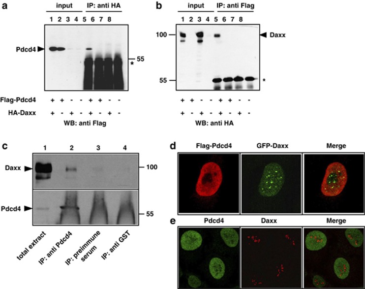
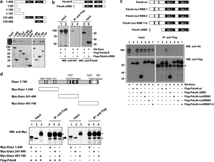
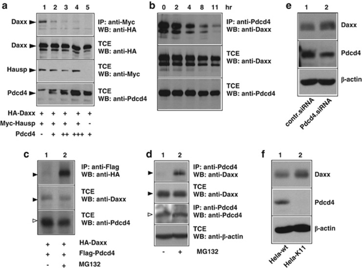
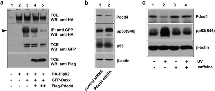
The tumor suppressor protein Pdcd4 is a nuclear/cytoplasmic shuttling protein that has been implicated in the development of several types of human cancer. In the nucleus, Pdcd4 affects the transcription of specific genes by modulating the activity of several transcription factors. We have identified the Daxx protein as a novel interaction partner of Pdcd4. Daxx is a scaffold protein with roles in diverse processes, including transcriptional regulation, DNA-damage signaling, apoptosis and chromatin remodeling. We show that the interaction of both proteins is mediated by the N-terminal domain of Pdcd4 and the central part of Daxx, and that binding to Pdcd4 stimulates the degradation of Daxx, presumably by disrupting the interaction of Daxx with the de-ubiquitinylating enzyme Hausp. Daxx has previously been shown to serve as a scaffold for protein kinase Hipk2 and tumor suppressor protein p53 and to stimulate the phosphorylation of p53 at serine 46 (Ser-46) in response to genotoxic stress. We show that Pdcd4 also disrupts the Daxx-Hipk2 interaction and inhibits the phosphorylation of p53. We also show that ultraviolet irradiation decreases the expression of Pdcd4. Taken together, our results support a model in which Pdcd4 serves to suppress the phosphorylation of p53 in the absence of DNA damage, while the suppressive effect of Pdcd4 is abrogated after DNA damage owing to the decrease of Pdcd4. Overall, our data demonstrate that Pdcd4 is a novel modulator of Daxx function and provide evidence for a role of Pdcd4 in restraining p53 activity in unstressed cells.


Figures

Figure 1
Figure 1
Co-immunoprecipitation and subnuclear localization of Pdcd4 and Daxx. (a, b) QT6 cells were transfected with the indicated combinations of plasmids encoding Flag-Pdcd4 and HA-Daxx. Cells were lysed after 24 h and protein extracts were immunoprecipitated with anti-Flag or anti-HA antibodies, as indicated above the panels. Proteins were analyzed by SDS–PAGE and western blotting, using the antibodies indicated below the panels. Analyses of the crude protein extracts (input) demonstrate comparable expression levels of the proteins in the different samples. HA-Daxx and Flag-Pdcd4 are marked by arrowheads. The asterisks mark the immunoglobulin heavy chains of the HA and Flag antibodies. (c) Protein extracts of HeLa cells were immunoprecipitated with an antiserum against endogenous human Pdcd4 (lane 2). Controls were performed with preimmune serum from the same animal (lane 3) or with an antiserum against tubulin (lane 4). Total cell extract (lane 1) and precipitated proteins were analyzed by SDS–PAGE, followed by western blotting using an antiserum against Daxx (upper panel) or Pdcd4 (bottom panel). Daxx and Pdcd4 are marked by black arrowheads. The strong diffuse staining in lanes 2–4 at the bottom of the lower panel is due to the immunoglobulins from the antiserum used for immunoprecipitation. (d) HeLa cells were transfected with expression vectors for Flag-Pdcd4 and GFP-Daxx. After 24 h, cells were fixed and Flag-Pdcd4 was stained with anti-Flag and tetramethyl rhodamine iso-thiocyanate-conjugated secondary antibody (red). GFP-Daxx was detected using intrinsic GFP fluorescence (green). (e) Non-transfected HeLa cells were stained with antiserum against endogenous Pdcd4 (green) and endogenous Daxx (red).
Figure 2
Figure 2
Mapping of the Daxx and Pdcd4 binding sites. (a) GST-pull down experiments were performed with the indicated GST and GST-Pdcd4 fusion proteins, which were incubated with lysates of QT6 cells transfected with an expression vector for HA-Daxx. Bound proteins were analyzed by SDS–PAGE, followed by western blotting with antibodies against the HA-tag. Total protein extract of the transfected cells was used as control (input). Coomassie blue staining of GST proteins used in the pull-down experiments demonstrates comparable protein amounts (lower panels). (b, c) QT6 cells were transfected with the indicated combinations of plasmids encoding full-length Flag-Pdcd4, N terminally truncated or point-mutated versions of Flag-Pdcd4, and HA-Daxx. Cells were lysed after 24 h and protein extracts were immunoprecipitated with anti-Flag antibodies, followed by SDS–PAGE and western blotting, using the indicated antibodies. Analyses of the crude protein extracts (input lanes) demonstrate comparable expression levels of the proteins in the different samples. The asterisk in panel c marks the immunoglobulin heavy chain of the Flag antibody. The Pdcd4 constructs used are shown schematically at the top of panels a–c. (d) QT6 cells were transfected with the indicated combinations of plasmids encoding full-length Flag-Pdcd4 and Myc-tagged Daxx proteins. Cells were lysed after 24 h and protein extracts were immunoprecipitated with anti-Flag antibodies, followed by SDS–PAGE and western blotting with anti-Myc antibodies. Aliquots of the crude protein extracts (input lanes) were analyzed to demonstrate the expression levels and the sizes of the Daxx constructs. The Myc-Daxx (241–490) protein is marked by an arrowhead. Asterisks mark the immunoglobulin heavy chain of the Flag antibody and a nonspecific protein band present in all immunoprecipitates. The Daxx constructs used are shown schematically at the top.
Figure 3
Figure 3
Pdcd4 disrupts the interaction of Daxx and Hausp and decreases the half-life of Daxx. (a) QT6 cells were transfected with the indicated combinations of expression vectors for HA-Daxx, Myc-Hausp and Pdcd4, as indicated below the lanes. Cells were lysed after 24 h and protein extracts were either analyzed directly by western blotting (panels labeled TCE (total protein extract)) or were first immunoprecipitated with antibodies against the HA-tag before western blot analysis (top panel). (b) QT6 cells were transfected with expression vectors for HA-Daxx and Flag-Pdcd4. At 24 h after transfection, 50 μg/ml cycloheximide was added to the growth medium and the cells were harvested immediately or after growing them for additional times, as indicated at the top. Cell extracts were immunoprecipitated with anti-Flag antibodies, followed by SDS–PAGE and western blotting with anti-HA antibodies (upper panel). Aliquots of the TCEs were analyzed with the indicated antibodies to demonstrate the Daxx and Pdcd4 expression levels (lower panels). (c) QT6 cells were transfected with expression vectors for HA-Daxx and Flag-Pdcd4. The cells were incubated with or without 10 nℳ MG132 for 4 h before they were lysed and immunoprecipitated with anti-Flag antibodies, followed by SDS–PAGE and western blotting with anti HA antibodies (upper panel). Aliquots of the TCEs were analyzed with the indicated antibodies to demonstrate the total expression levels of the proteins (lower panels). (d) HeLa cells were incubated with or without 10 nℳ MG132 for 4 h before they were lysed. Cell extracts were then immunoprecipitated with anti-Pdcd4 antibodies, followed by SDS–PAGE and western blotting with anti-Daxx antibodies (upper panel). Aliquots of the TCEs were analyzed with the indicated antibodies to demonstrate the expression levels of endogenous Daxx, Pdcd4 and β-actin (lower panels). To demonstrate the MG132-dependent increase of co-precipitated transfected or endogenous Daxx, the upper panels of (c) and (d) were exposed for a short time only. Daxx co-precipitated from cells not treated with MG132 is therefore only weakly visible. (e) MCF7 cells were transfected with control siRNA or Pdcd4-specific siRNA. The cells were analyzed after 2 days by western blotting for the expression of Daxx, Pdcd4 and β-actin. (f) HeLa wild-type cells or a clone of HeLa cells stably expressing Pdcd4-specific short hairpin RNA (HeLa-K11) were analyzed as described in (e).
Figure 4
Figure 4
Pdcd4 inhibits Ser-46 phosphorylation of p53. (a) QT6 cells were transfected with the indicated combinations of expression vectors for HA-Hipk2, GFP-Daxx and Flag-Pdcd4, as indicated below the lanes. Cells were lysed after 24 h and TCEs were either analyzed directly by SDS–PAGE and western blotting with the indicated antibodies or were first immunoprecipitated with antibodies against GFP (second panel from top) before western blot analysis. Hipk2 co-precipitated via Daxx is marked by an arrowhead. (b) MCF7 cells were treated with Pdcd4-specific or control siRNA for 48 h. TCEs were subsequently analyzed with antibodies against Pdcd4, p53 and β-actin. In addition, p53 was first immunoprecipitated from the cell extracts and the immunoprecipitates were then analyzed by western blotting with phospho-p53- (Ser-46) specific antibodies. (c) HEK293 cells were UV irradiated (50 J/cm2) in the presence or absence of caffeine (concentration 6 mℳ). Unirradiated cells served as control. At 4 h after irradiation, TCEs were analyzed by western blotting with antibodies against Pdcd4, phospho-p53(Ser-46) and β-actin.

References

    1. Shibahara K, Asano M, Ishida Y, Aoki T, Koike T, Honjo T. Isolation of a novel mouse gene MA-3 that is induced upon programmed cell death. Gene. 1995;166:297–301. - PubMed
    1. Cmarik JL, Min H, Hegamyer G, Zhan S, Kulesz-Martin M, Yoshinaga H, et al. Differentially expressed protein Pdcd4 inhibits tumor promoter-induced neoplastic transformation. Proc Natl Acad Sci USA. 1999;96:14037–14042. - PMC - PubMed
    1. Jansen AP, Camalier CE, Colburn NH. Epidermal expression of the translation inhibitor programmed cell death 4 suppresses tumorigenesis. Cancer Res. 2005;65:6034–6041. - PubMed
    1. Chen Y, Knosel T, Kristiansen G, Pietas A, Garber ME, Matsuhashi S, et al. Loss of PDCD4 expression in human lung cancer correlates with tumour progression and prognosis. J Pathol. 2003;200:640–646. - PubMed
    1. Afonja O, Juste D, Das S, Matsuhashi S, Samuels HH. Induction of PDCD4 tumor suppressor gene expression by RAR agonists, antiestrogen and HER-2/neu antagonist in breast cancer cells. Evidence for a role in apoptosis. Oncogene. 2004;23:8135–8145. - PubMed