. 2013 Mar 27;5(178):178ra40.
doi: 10.1126/scitranslmed.3005580.

NETs are a source of citrullinated autoantigens and stimulate inflammatory responses in rheumatoid arthritis

Affiliations

NETs are a source of citrullinated autoantigens and stimulate inflammatory responses in rheumatoid arthritis

Ritika Khandpur et al. Sci Transl Med. .

Abstract

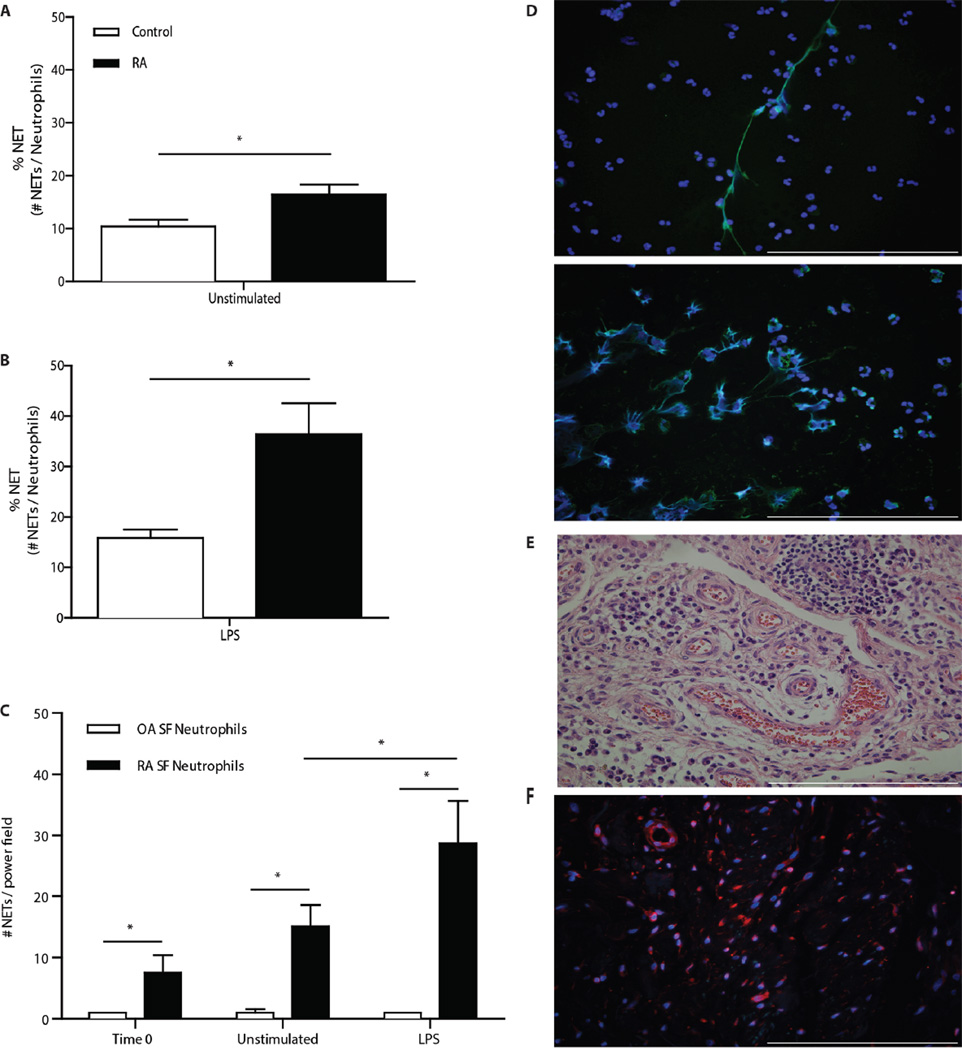
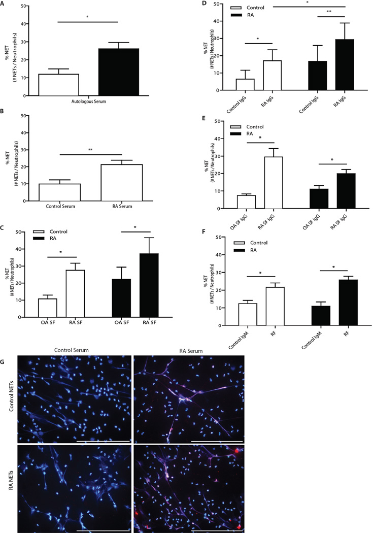
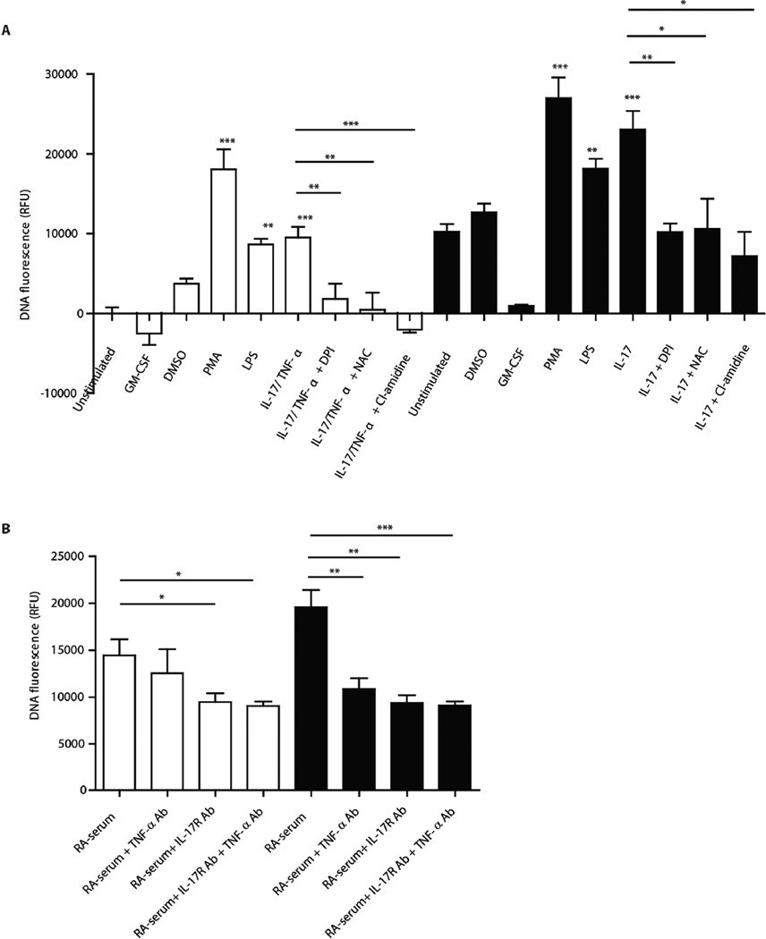
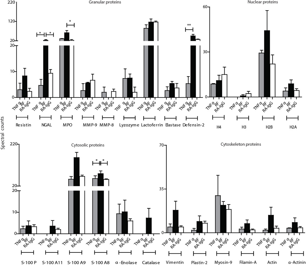
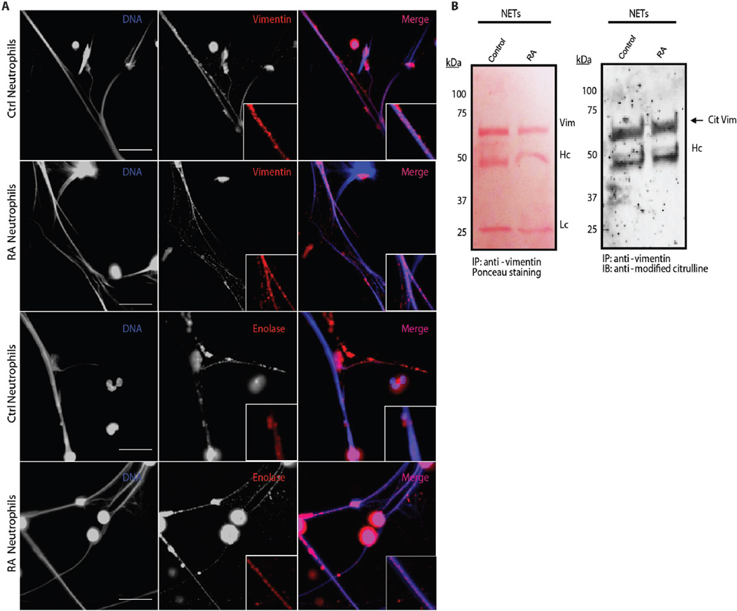
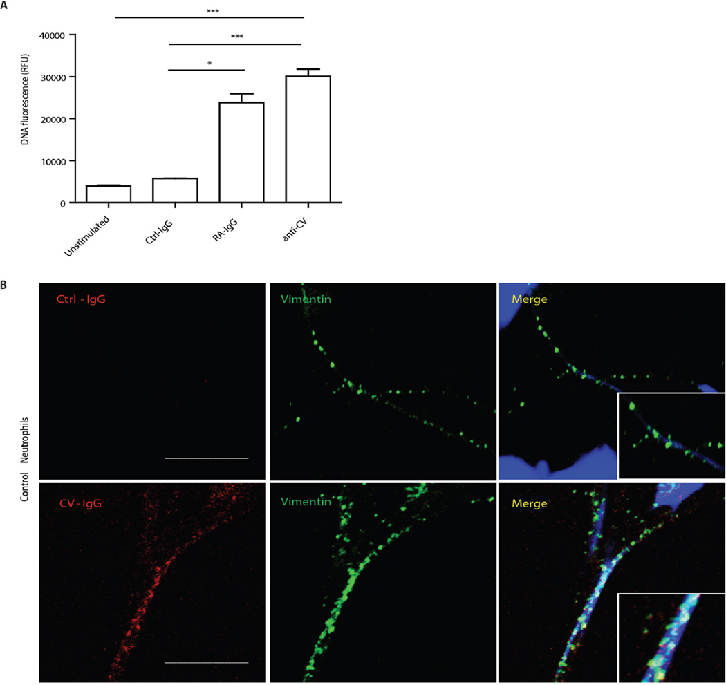
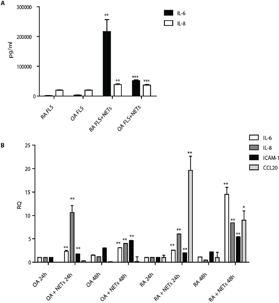
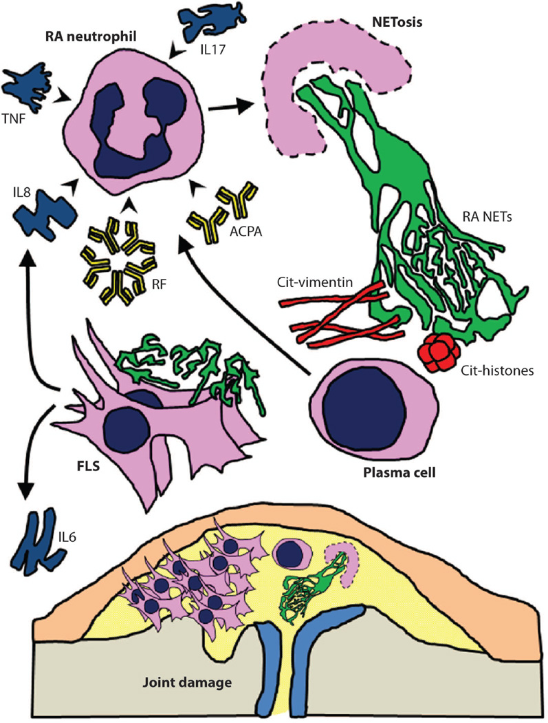
The early events leading to the development of rheumatoid arthritis (RA) remain unclear, but formation of autoantibodies to citrullinated protein antigens (ACPAs) is considered a key pathogenic event. Neutrophils isolated from patients with various autoimmune diseases display enhanced neutrophil extracellular trap (NET) formation, a phenomenon that exposes autoantigens in the context of immunostimulatory molecules. We investigated whether aberrant NETosis occurs in RA, determined its triggers, and examined its deleterious inflammatory consequences. Enhanced NETosis was observed in circulating and RA synovial fluid neutrophils compared to neutrophils from healthy controls and from patients with osteoarthritis (OA). Further, netting neutrophils infiltrated RA synovial tissue, rheumatoid nodules, and skin. NETosis correlated with ACPA presence and levels and with systemic inflammatory markers. RA sera and immunoglobulin fractions from RA patients with high levels of ACPA and/or rheumatoid factor significantly enhanced NETosis, and the NETs induced by these autoantibodies displayed distinct protein content. Indeed, during NETosis, neutrophils externalized the citrullinated autoantigens implicated in RA pathogenesis, and anti-citrullinated vimentin antibodies potently induced NET formation. Moreover, the inflammatory cytokines interleukin-17A (IL-17A) and tumor necrosis factor-α (TNF-α) induced NETosis in RA neutrophils. In turn, NETs significantly augmented inflammatory responses in RA and OA synovial fibroblasts, including induction of IL-6, IL-8, chemokines, and adhesion molecules. These observations implicate accelerated NETosis in RA pathogenesis, through externalization of citrullinated autoantigens and immunostimulatory molecules that may promote aberrant adaptive and innate immune responses in the joint and in the periphery, and perpetuate pathogenic mechanisms in this disease.


Figures

Figure 1
Figure 1. Enhanced NETosis in RA neutrophils
A and B. Peripheral blood (PB) RA neutrophils undergo increased NETosis in the absence (A) or presence (B) of added stimulation (LPS for 1 h); (control, n=7; RA, n=13–14). C. SF RA neutrophils undergo increased NETosis in the absence (unstimulated) or presence of 1 h LPS stimulation, n=5/group, *p<0.05. Results represent mean ± SEM D. Representative microphotographs display enhanced LPS-induced NETosis in RA (bottom) vs. control PB neutrophils (top). NETs were visualized as structures co-staining for neutrophil elastase (green) and DAPI (blue); 40x magnification. E. and F. Multilobate inflammatory cells consistent with netting neutrophils are present in RA synovial tissue. E represents H&E stain (400X) of RA synovial tissue. F represents same area stained with anti-MPO (red) and DAPI (blue). Squares represent areas where netting neutrophils were observed. Images are representative of 3 RA patients. Statistical analysis was with two-tailed unpaired t tests except for SF experiments where paired t-test was used. Bar is 10 µm.
Figure 2
Figure 2. AutoAbs present in RA serum and SF induce NETosis and bind to NETs
A–C: RA serum and SF induce significantly higher NETosis in RA autologous neutrophils (white bar) and control neutrophils (black bar), when compared to the effect of control sera or OA SF on autologous neutrophils or RA neutrophils. D–E. Similar effects were observed when comparing IgG purified from RA serum or from SF from patients with seropositive RA, compared to control serum or OA SF. F. Purified IgM RF significantly enhances NETs in RA and control neutrophils (n=3–8/group). The % of NETosis (elastase and DAPI labeled neutrophils/total neutrophils) was quantified after 1 h exposure to serum or SF. Results represent mean ± SEM; *p<0.05; **p<0.01 using two-tailed unpaired t tests. G. RA IgG isolated from patients with higher titer ACPA and/or RF binds to RA and control NETs induced by LPS. Results are representative of 3 independent experiments. Bar is 10 µm. NC: normal control; OA: osteoarthritis: RA: rheumatoid arthritis, SF: synovial fluid; RF: rheumatoid factor.
Figure 3
Figure 3. Proinflammatory cytokines induce NETosis and this is enhanced in RA neutrophils
A. Results are adjusted to levels observed in unstimulated control neutrophils. DPI: diphenyleneiodonium chloride; NAC: N-acetyl cysteine; Cl-amidine: PAD inhibitor. B. Neutralizing Abs to TNF and/or IL-17R decrease NETosis induced by RA sera. Results are expressed as relative fluorescent units (RFUs) of DNA fluorescence using Sytox green assay. Results represent mean ±SEM of 3 independent experiments, performed in triplicate; * p<0.05, **p < 0.005, ***p < 0.0001, using two-tailed (A) and one-tailed (B) unpaired t tests.
Figure 4
Figure 4. Various stimuli present in RA sera induce distinct protein cargo in control NETs
Bar graphs represent spectral counts from 25 of the proteins identified in the NETs, which were normalized for protein content. RF=rheumatoid factor, RA IgG=IgG isolated from RA patients with high titers of ACPA and/or RF; *p<0.05, ** p<0.01, when comparing proteins expressed in the NETs induced by the 3 conditions. Results represent mean ±SEM of 3–5 independent experiments. Comparisons among groups were done with one-way ANOVA and p values were adjusted for Bonferroni’s multiple comparison test.
Figure 5
Figure 5
A. Vimentin and α-enolase decorate control and RA NETs. Red represents vimentin or α-enolase and blue is Hoechst. Total magnification 63X. B. Vimentin externalized in control and RA NETs is citrullinated. Left panel is visualization with Ponceau. Right panel depicts immunoblot using anti-modified citrulline and detection by chemiluminescence (right). Molecular weight markers (kDa) are indicated. Cit Vim=citrullinated vimentin; Hc=heavy chain; Lc=light chain; Vim=vimentin; IP: immunoprecipitate; IB: Immunoblot. Results are representative of 3 independent experiments. Bar is 10 µm.
Figure 6
Figure 6
A. Anti-CV Abs isolated from RA sera significantly enhance control neutrophil NETosis. Results represent mean+SEM of 3 independent experiments, each performed in duplicate. Units are expressed as RFUs of DNA fluorescence using Sytox green assay; *p<0.05, ***p<0.001, using two-tailed unpaired t test. B. Anti-CV Abs bind to vimentin externalized in the NETs. Original magnification is 63x; Ctrl=control. Results are representative of 2 independent experiments. Bar is 10 µm.
Figure 7
Figure 7
A. RA and OA FLS upregulate inflammatory cytokine synthesis upon exposure to RA NETs for 48 h. Results represent mean±SEM of 2 independent experiments, each performed in triplicate-quadruplicate; *p<0.05, ** p<0.01, ***p<0.001, using two-tailed unpaired t tests. B. RA and OA FLS upregulate mRNA synthesis of inflammatory cytokines, chemokines and adhesion molecules upon exposure to RA NETs. Results are expressed as fold induction of the specific gene at 48 h, when compared to untreated FLS at 24 h of culture, adjusted for housekeeping gene (GAPDH). Error bars represent maximum RQ value for each sample and represent mean±SEM of 2–3 independent experiments. *p<0.05 comparing change in mean DCt compared with untreated samples at same time-point, using two-tailed unpaired t tests. For untreated OA FLS, there was no amplification of CCL20, suggesting the basal expression was below the level of detection.
Figure 8
Figure 8. Proposed model of the role of NETosis in RA
RA NETs may provide a source of autoAgs and activate FLS and B cells. AutoAbs (RF, ACPA) and inflammatory cytokines (IL-17A, TNF-α, and IL-8) are all potential stimuli for enhanced NETosis in RA. In turn, RA NETs are a source of citrullinated (Cit-) autoAgs, including vimentin, further triggering production of ACPA. RA NETs also promote activation and cytokine release by FLS, with implications for joint damage and further propagation of a vicious cycle of NET induction and autoAb formation.

Comment in

References

    1. Tarner IH, Muller-Ladner U, Gay S. Emerging targets of biologic therapies for rheumatoid arthritis. Nat Clin Pract Rheumatol. 2007;3:336–345. - PubMed
    1. Deane KD, Norris JM, Holers VM. Preclinical rheumatoid arthritis: identification evaluation and future directions for investigation. Rheum Dis Clin North Am. 2010;36:213–241. - PMC - PubMed
    1. Makrygiannakis D, af Klint E, Lundberg IE, Lofberg R, Ulfgren AK, Klareskog L, Catrina AI. Citrullination is an inflammation-dependent process. Ann Rheum Dis. 2006;65:1219–1222. - PMC - PubMed
    1. Kidd BA, Ho PP, Sharpe O, Zhao X, Tomooka BH, Kanter JL, Steinman L, Robinson WH. Epitope spreading to citrullinated antigens in mouse models of autoimmune arthritis and demyelination. Arthritis Res Ther. 2008;10:R119. - PMC - PubMed
    1. Lundberg K, Nijenhuis S, Vossenaar ER, Palmblad K, van Venrooij WJ, Klareskog L, Zendman AJ, Harris HE. Citrullinated proteins have increased immunogenicity and arthritogenicity and their presence in arthritic joints correlates with disease severity. Arthritis Res Ther. 2005;7:R458–R467. - PMC - PubMed

Publication types

MeSH terms