. 2013 Mar 27;33(13):5524-32.
doi: 10.1523/JNEUROSCI.4384-12.2013.

ACR-12 ionotropic acetylcholine receptor complexes regulate inhibitory motor neuron activity in Caenorhabditis elegans

Affiliations

ACR-12 ionotropic acetylcholine receptor complexes regulate inhibitory motor neuron activity in Caenorhabditis elegans

Hilary A Petrash et al. J Neurosci. .

Abstract

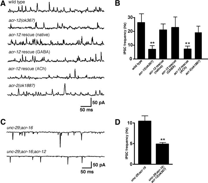
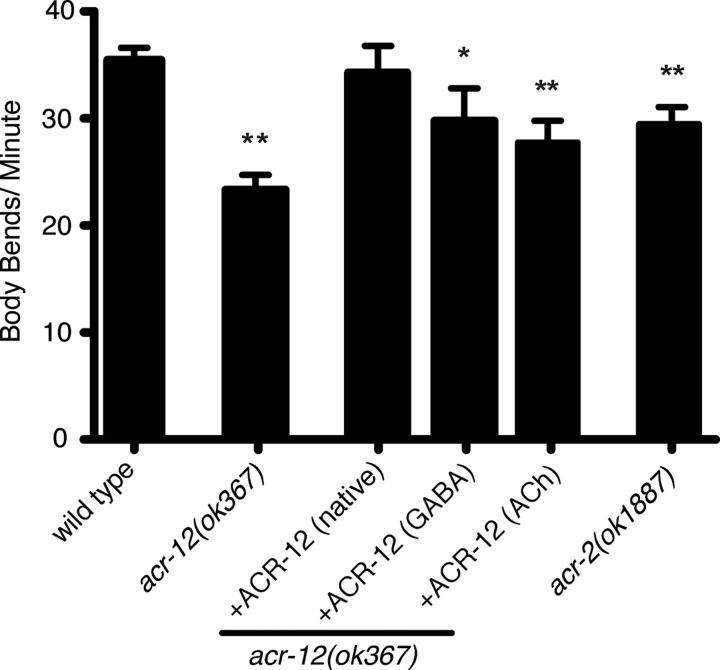
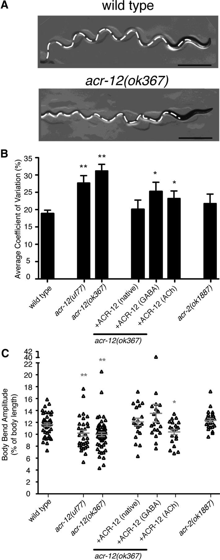
Heterogeneity in the composition of neurotransmitter receptors is thought to provide functional diversity that may be important in patterning neural activity and shaping behavior (Dani and Bertrand, 2007; Sassoè-Pognetto, 2011). However, this idea has remained difficult to evaluate directly because of the complexity of neuronal connectivity patterns and uncertainty about the molecular composition of specific receptor types in vivo. Here we dissect how molecular diversity across receptor types contributes to the coordinated activity of excitatory and inhibitory motor neurons in the nematode Caenorhabditis elegans. We show that excitatory and inhibitory motor neurons express distinct populations of ionotropic acetylcholine receptors (iAChRs) requiring the ACR-12 subunit. The activity level of excitatory motor neurons is influenced through activation of nonsynaptic iAChRs (Jospin et al., 2009; Barbagallo et al., 2010). In contrast, synaptic coupling of excitatory and inhibitory motor neurons is achieved through a second population of iAChRs specifically localized at postsynaptic sites on inhibitory motor neurons. Loss of ACR-12 iAChRs from inhibitory motor neurons leads to reduced synaptic drive, decreased inhibitory neuromuscular signaling, and variability in the sinusoidal motor pattern. Our results provide new insights into mechanisms that establish appropriately balanced excitation and inhibition in the generation of a rhythmic motor behavior and reveal functionally diverse roles for iAChR-mediated signaling in this process.


Figures

Figure 1.
Figure 1.
ACR-12 gene sequence. A, ACR-12 amino acid sequence. cys-loop (*), vicinal cysteines present in α subunits (∼), GFP insertion site (green lettering), transmembrane domains (gray shading), ok367 deletion (blue lettering), and site of uf77 mutation (yellow shading) are indicated. B, Schematic of ACR-12 receptor subunit. Approximate locations of cys-loop (*), vicinal cysteines (∧∧), GFP insertion site (green box), transmembrane domains (M1–M4), ok367 deletion (red shading), and uf77 mutation (yellow star) are indicated. C, Time course of paralysis in the presence of aldicarb (1 mm) for wild-type (blue) (n = 16), acr-12(ok367) mutants (red) (n = 14), acr-12(uf77) mutants (gray) (n = 8), unc-49(e382) mutants (brown) (n = 11), unc-49;acr-12(ok367) double mutants (green) (n = 10), ACR-12–GFP rescue using the native promoter (purple) (n = 12), and acr-2(ok1887) mutants (black) (n = 4) are shown. Data represent mean ± SEM.
Figure 2.
Figure 2.
ACR-12 iAChRs regulate GABA MN activity. A, Diagram of C. elegans showing ventral nerve cord. Red box indicates approximate region imaged in B and C. B, Confocal image showing coexpression of ACR-12–GFP (ufIs38) and a ACh MN-specific marker (Pacr-2::mCherry, ufIs43) in the posterior ventral nerve cord. For B and C, asterisks indicate MN cell bodies coexpressing both reporters. Scale bars, 50 μm. C, Confocal image showing coexpression of ACR-12–GFP (ufIs38) and a GABA neuron-specific marker (Punc-47::mCherry, ufIs34) in the posterior ventral nerve cord. D, Time course of paralysis in the presence of aldicarb (1 mm) for wild-type (blue) (n = 14), acr-12(ok367) mutants (red) (n = 14), ACh-specific (Pacr-2) ACR-12–GFP rescue (purple) (n = 10), and GABA-specific (Punc-47) ACR-12–GFP rescue (black) (n = 10).
Figure 3.
Figure 3.
ACR-12 is differentially localized across MN populations. A, Schematic of C. elegans locomotory circuit. Red coloring indicates ACh MNs (VA, VB, DA, and DB) and blue indicates GABA MNs (DD and VD). Dark gray and light gray shading represent body wall muscles. In A and C, black triangles and boxes represent sites of cholinergic (excitatory) and GABAergic (inhibitory) innervation of muscles, respectively. Figure modified from WormAtlas (http://www.wormatlas.org/). B, Independent confocal images showing ACR-12–GFP localization in ventral and dorsal nerve cords of young adult animals as indicated (ufIs57). Positions of cell bodies are indicated by white arrowheads in B, D, and E. Scale bars, 10 μm. C, Diagram depicting subcellular localization of ACR-12–GFP and the presynaptic marker mCherry–RAB-3 in ACh and GABA MNs. Although ACh MNs form dyadic synapses onto both GABA MNs and muscles, only synaptic contacts between ACh and GABA MNs are depicted for clarity. ACR-12–GFP fluorescence (green shading) is diffuse in the processes of ACh VA and VB MNs. In contrast, ACR-12–GFP fluorescence appears punctate (green circles) in the processes of GABAergic VD and DD MNs and localizes opposite mCherry–RAB-3 fluorescent puncta (red circles) expressed in ACh MNs. D, Confocal image of Pacr-2::ACR-12–GFP (ufIs78) localization in ACh MNs of the posterior ventral nerve cord. E, Confocal images of Punc-47::ACR-12–GFP (ufIs92) localization in GABA MNs of the posterior ventral and dorsal nerve cords as indicated. F, Confocal image of the dorsal nerve cord showing apposition of mCherry–RAB-3 expressed in ACh MNs (Pacr-2, ufIs63) with ACR-12–GFP expressed in GABA MNs (Punc-47, ufIs92). Scale bar, 10 μm.
Figure 4.
Figure 4.
EPSC frequency is reduced in acr-12 mutants. A, Representative recordings of endogenous EPSCs recorded at −60 mV for the indicated genotypes. Rescue refers to transgenic expression of ACR-12 in acr-12 mutants using the native promoter. B, Average (mean ± SEM) EPSC frequency for the genotypes indicated. **p < 0.01 from wild type. Wild type, n = 11; acr-12, n = 13; rescue, n = 6.
Figure 5.
Figure 5.
Loss of ACR-12 receptors from GABA MNs reduces inhibitory signaling. A, Representative recordings of endogenous IPSCs recorded at 0 mV for the indicated genotypes. Rescue refers to transgenic expression of ACR-12 in acr-12 mutants under control of either the native, unc-47 (GABA) or acr-2 (ACh) promoters. B, Average (mean ± SEM) endogenous IPSC frequency for the genotypes indicated. Wild type, n = 7; acr-12, n = 9; acr-12 rescue (native), n = 4; acr-12 rescue (GABA), n = 5, acr-12 rescue (ACh), n = 4; acr-2, n = 4. **p < 0.01 from wild type. C, Representative recordings of endogenous IPSCs recorded at −60 mV for the indicated genotypes. Recordings were made using 115 mm KCl in the pipette. D, Average (mean ± SEM) endogenous IPSC frequency for the genotypes indicated. unc-29;acr-16, n = 12; unc-29;acr-16;acr-12, n = 11. **p < 0.01 from unc-29;acr-16.
Figure 6.
Figure 6.
ACR-12 iAChRs regulate the velocity of movement. Average (mean ± SEM) number of body bends per minute for the genotypes indicated. Wild type, n = 36; acr-12(ok367), n = 29; ACR-12–GFP rescue (native promoter), n = 10; GABA-specific (Punc-47) ACR-12–GFP rescue, n = 10; ACh-specific (Pacr-2) ACR-12–GFP rescue, n = 10; and acr-2(ok1887), n = 17. *p < 0.05 from wild type; **p < 0.01 from wild type.
Figure 7.
Figure 7.
ACR-12 iAChRs are required for consistent motor pattern. A, Still images of locomotory paths for wild-type and acr-12(ok367) mutants as indicated. Tracks are indicated by white dashed lines. The image shows a more severe example of the variable waveform phenotype in acr-12 mutants. Scale bar, 2 mm. B, Coefficient of variance for body bend amplitude averaged over 10 consecutive body bends for genotypes indicated. Wild type, n = 23; acr-12(uf77), n = 11; acr-12(ok367), n = 52; rescue (native promoter), n = 11; rescue (GABA-specific), n = 34; rescue (ACh-specific), n = 14; acr-2(ok1887), n = 13. *p < 0.05 from wild type; **p < 0.01 from wild type. C, Scatter plot of body bend amplitudes for genotypes indicated. Wild type, n = 45; acr-12(uf77), n = 31; acr-12(ok367), n = 52; rescue (native), n = 20; rescue (GABA-specific), n = 20; rescue (ACh-specific), n = 20; acr-2(ok1887), n = 24. Each point represents an average of three consecutive body bends for an individual animal. Mean ± SEM is indicated by horizontal lines. *p < 0.05 from WT; **p < 0.01 from wild type.

References

    1. Bamber BA, Beg AA, Twyman RE, Jorgensen EM. The Caenorhabditis elegans unc-49 locus encodes multiple subunits of a heteromultimeric GABA receptor. J Neurosci. 1999;19:5348–5359. - PMC - PubMed
    1. Bany IA, Dong MQ, Koelle MR. Genetic and cellular basis for acetylcholine inhibition of Caenorhabditis elegans egg-laying behavior. J Neurosci. 2003;23:8060–8069. - PMC - PubMed
    1. Barbagallo B, Prescott HA, Boyle P, Climer J, Francis MM. A dominant mutation in a neuronal acetylcholine receptor subunit leads to motor neuron degeneration in Caenorhabditis elegans. J Neurosci. 2010;30:13932–13942. - PMC - PubMed
    1. Boulin T, Gielen M, Richmond JE, Williams DC, Paoletti P, Bessereau JL. Eight genes are required for functional reconstitution of the Caenorhabditis elegans levamisole-sensitive acetylcholine receptor. Proc Natl Acad Sci U S A. 2008;105:18590–18595. - PMC - PubMed
    1. Cinar H, Keles S, Jin Y. Expression profiling of GABAergic motor neurons in Caenorhabditis elegans. Curr Biol. 2005;15:340–346. - PubMed

Publication types

MeSH terms

LinkOut - more resources