. 2013 May 10;288(19):13522-33.
doi: 10.1074/jbc.M113.455329. Epub 2013 Mar 27.

Alternative C-terminal helix orientation alters chemokine function: structure of the anti-angiogenic chemokine, CXCL4L1

Affiliations

Alternative C-terminal helix orientation alters chemokine function: structure of the anti-angiogenic chemokine, CXCL4L1

Je-Hung Kuo et al. J Biol Chem. .

Abstract

Background: CXCL4L1 is a highly potent anti-angiogenic and anti-tumor chemokine, and its structural information is unknown.

Results: CXCL4L1 x-ray structure is determined, and it reveals a previously unrecognized chemokine structure adopting a novel C-terminal helix conformation.

Conclusion: The alternative helix conformation enhances the anti-angiogenic activity of CXCL4L1 by reducing the glycosaminoglycan binding ability.

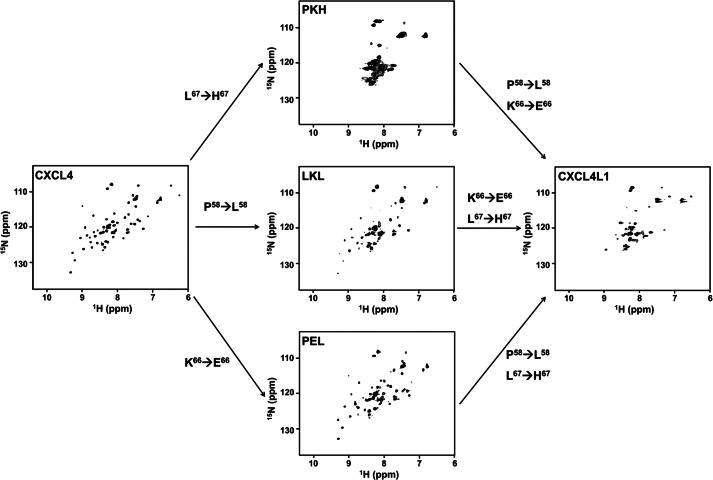
Significance: Chemokine C-terminal helix orientation is critical in regulating their functions. Chemokines, a subfamily of cytokines, are small, secreted proteins that mediate a variety of biological processes. Various chemokines adopt remarkable conserved tertiary structure comprising an anti-parallel β-sheet core domain followed by a C-terminal helix that packs onto the β-sheet. The conserved structural feature has been considered critical for chemokine function, including binding to cell surface receptor. The recently isolated variant, CXCL4L1, is a homologue of CXCL4 chemokine (or platelet factor 4) with potent anti-angiogenic activity and differed only in three amino acid residues of P58L, K66E, and L67H. In this study we show by x-ray structural determination that CXCL4L1 adopts a previously unrecognized structure at its C terminus. The orientation of the C-terminal helix protrudes into the aqueous space to expose the entire helix. The alternative helix orientation modifies the overall chemokine shape and surface properties. The L67H mutation is mainly responsible for the swing-out effect of the helix, whereas mutations of P58L and K66E only act secondarily. This is the first observation that reports an open conformation of the C-terminal helix in a chemokine. This change leads to a decrease of its glycosaminoglycan binding properties and to an enhancement of its anti-angiogenic and anti-tumor effects. This unique structure is recent in evolution and has allowed CXCL4L1 to gain novel functional properties.

Keywords: Angiogenesis; CXCL4L1; Chemokines; Heparin; Platelet Factor 4; Platelets; X-ray Crystallography.


Figures

FIGURE 1.
FIGURE 1.
Schematic representation of the amino acid sequences of CXCL4, CXCL4L1, and the variants. The substituted residues in the mutants are highlighted in bold. The secondary structural elements determined by x-ray crystallography are indicated at the top of the sequence. Two connecting lines between Cys residues represent the disulfide connectivities of Cys-10 to Cys-36 and Cys-12 to Cys-52.
FIGURE 2.
FIGURE 2.
The structure, oligomerization state, and heparin binding properties of DTT-reduced CXCL4. A, 1H,15N HSQC spectra of CXCL4 (left) and DTT-reduced CXCL4 (right) at 600 MHz and 30 °C are shown. Two sets of resonances represent the molecular asymmetry of the tetramer (boxed, left). After reduction by the addition of 10 mm DTT, tetrameric CXCL4 with A, B, C, and D subunits was converted to an asymmetric A-B dimer. The heparin-binding sites, constituted by two C-terminal helices, are indicated by gray circles. B, shown is a secondary structure of DTT-reduced CXCL4, evaluated based on carbon chemical shifts (Δδ − Δδ). The values of Δδ and Δδ were, respectively, calculated from the differences between the experimental chemical shifts of 13Cα and 13Cβ and corresponding random coil values. The value of (Δδ − Δδ) for each residue represents the average of three consecutive residues (i − 1, i, i + 1), centered at residue i. Positive values are indicative of α-helices, and negative values represent β-strands. C, shown are far-UV CD spectra of CXCL4 and DTT-reduced CXCL4. D, shown is a FPLC size-exclusion chromatogram CXCL4 and DTT-reduced CXCL4. CXCL4 elutes at the position corresponding to tetramer, and DTT-reduced CXCL4 elutes at the position of an expected dimer. The elution positions of protein standards are indicated by arrows. E, heparin binding affinities of CXCL4 and DTT-reduced CXCL4 were analyzed by a Scatchard plot derived from SPR equilibrium responses at protein concentrations of 3–55 nm. The slopes of the best-fit linear correlations in the Scatchard plot represent the binding constant (KD) values of 3 and 91 nm for the CXCL4 tetramer and dimer, respectively. F, shown are competition ratios of SPR responses of CXCL4 and DTT-reduced CXCL4 in the addition of depolymerized (dp) heparin fragments. A protein concentration of 100 nm and heparin concentration of 10 μg/ml were mixed and injected.
FIGURE 3.
FIGURE 3.
Comparison of biophysical properties and heparin binding of CXCL4L1 and CXCL4. A, FPLC size-exclusion elution profiles are shown. The elution positions of protein standards are indicated by arrows. CXCL4 and CXCL4L1 both are eluted at the position corresponding to a tetramer. B, analytical ultracentrifugation (AUC) profiles are shown. The estimated apparent molecular masses are 28 and 32 kDa for CXCL4 and CXCL4L1, respectively. C, far-UV CD spectra for CXCL4 and CXCL4L1 are shown. D, shown are fluorescent emission spectra of ANS fluorescent dye after binding to the hydrophobic surface of CXCL4 and CXCL4L1. The stronger fluorescent intensity represents the larger portion of the hydrophobic surface exposed to the solvent. E, SPR sensograms of heparin binding are shown. Left, sensograms were obtained by injecting different concentrations of CXCL4 or CXCL4L1 (3–55 nm) over a heparin-coating chip. The inset shows the injections of CXCL4L1 with higher concentrations (0.3–1.5 μm), which were carried out due to very low affinity of CXCL4L1 for heparin. Right, sensograms were derived from injections of the same concentration (55 nm) of CXCL4, CXCL4L1, and the variants of LKL, PEL, PKH.
FIGURE 4.
FIGURE 4.
Crystal structure of CXCL4L1. A, shown is a schematic representation of CXCL4L1 tetramer (left) and the A-B and A-D interface where A/B/C/D monomer subunits are depicted by different colors. The hydrogen bonds for monomer association in A-B and A-D dimer interfaces are indicated by dotted lines. The residues constituting the exposed hydrophobic surface in A-B interface are shown as well as the residues responsible for association of N terminus in A-D interface. B, shown is the superposition of CXCL4 (PDB code 1F9Q) and CXCL4L1. The β-sheet core domain is almost identical, but helix α1 is rotated by ∼87° in CXCL4L1 relative to CXCL4, thus altering the overall shape of the molecule. The positions of the three mutations are indicated in two of the subunits by sticks. C, the distribution of positively charged residues and Glu-66 in CXCL4L1 is shown. D, the distribution of positively charged residues in CXCL4 is shown. The surrounding positive region is purposed to be able to recruit two individual anionic polysaccharides.
FIGURE 5.
FIGURE 5.
Orientations of helix α1 in CXCL4 and CXCL4L1. A, the two helical orientations (A/D and B/C) were identified in CXCL4L1 monomers, and only one orientation (A/B/C/D) was found in CXCL4 monomers. B, shown is a secondary structure for CXCL4 and CXCL4L1. Two disulfide bonds of Cys-10 to Cys-36 and Cys-12 to Cys-52 are shown as open curved bars. The hydrogen bonds of the backbone are indicated by arrows pointed from hydrogen bond donor to acceptor. The helix α1 contains residues Leu-59—Ser-70 in CXCL4 and residues Asp-54–His-67 in CXCL4L1, and the helical wheel of CXCL4 α1 is depicted to show the amphipathic property.
FIGURE 6.
FIGURE 6.
Effect of mutations on chemokine folding and stability. 1H,15N HSQC spectra of DTT-reduced CXCL4, CXCL4L1, and PKH, LKL, and PEL mutants are compared (0.2 mm protein concentration, 10 mm DTT, 25 °C, and 600 MHz).

Similar articles

Cited by

References

    1. Raman D., Sobolik-Delmaire T., Richmond A. (2011) Chemokines in health and disease. Exp. Cell Res. 317, 575–589 - PMC - PubMed
    1. Keeley E. C., Mehrad B., Strieter R. M. (2011) Chemokines as mediators of tumor angiogenesis and neovascularization. Exp. Cell Res. 317, 685–690 - PMC - PubMed
    1. Mukaida N., Baba T. (2012) Chemokines in tumor development and progression. Exp. Cell Res. 318, 95–102 - PubMed
    1. Bikfalvi A. (2012) Angiogenesis and invasion in cancer. Handb. Clin. Neurol. 104, 35–43 - PubMed
    1. Balkwill F. R. (2012) The chemokine system and cancer. J. Pathol. 226, 148–157 - PubMed

Publication types

MeSH terms

Associated data

LinkOut - more resources