. 2013 Apr 9;110(15):6067-72.
doi: 10.1073/pnas.1220572110. Epub 2013 Mar 27.

Eosinophil adoptive transfer system to directly evaluate pulmonary eosinophil trafficking in vivo

Affiliations

Eosinophil adoptive transfer system to directly evaluate pulmonary eosinophil trafficking in vivo

Ting Wen et al. Proc Natl Acad Sci U S A. .

Abstract

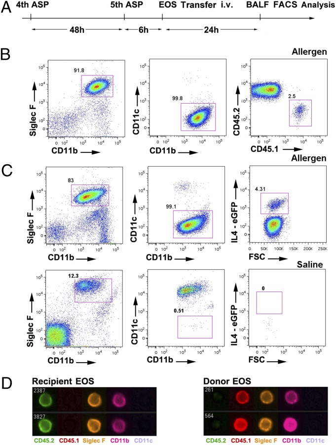
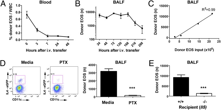
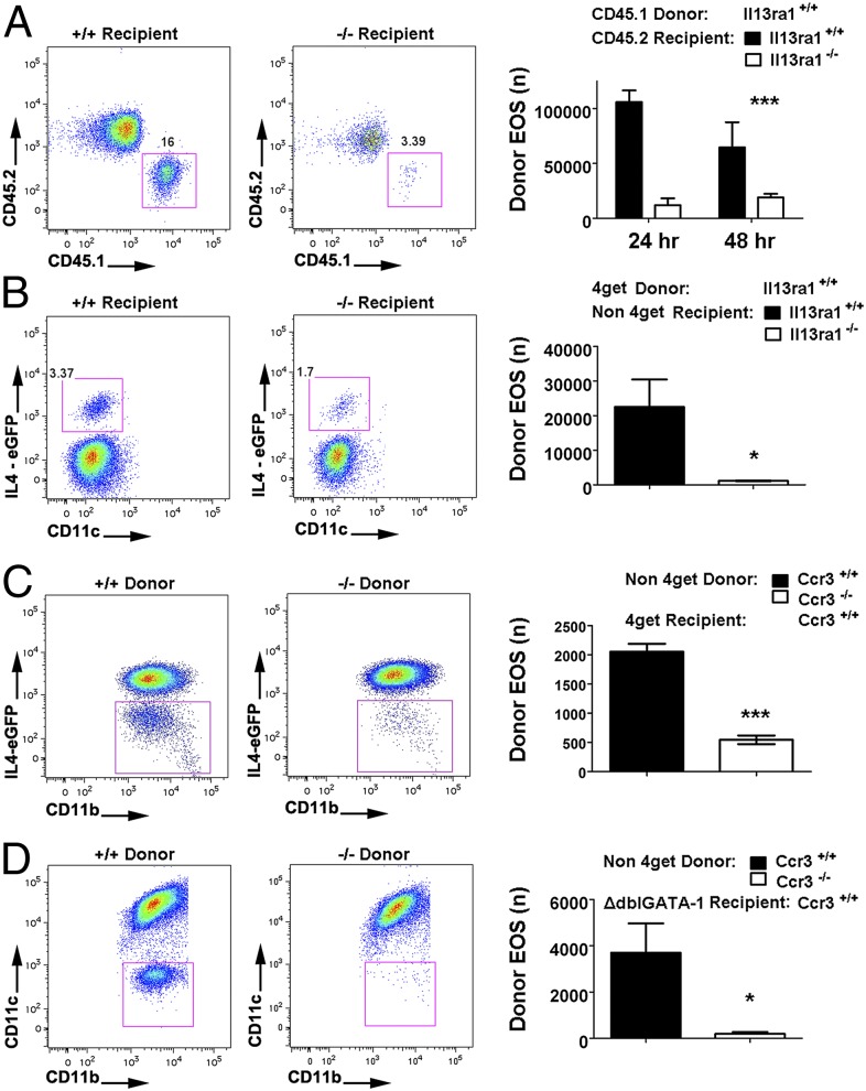
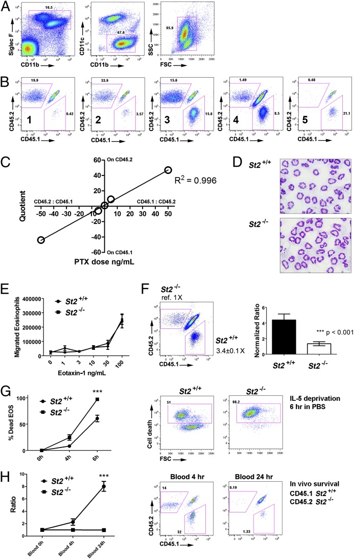
Most in vivo studies of granulocytes draw conclusions about their trafficking based on examination of their steady-state tissue/blood levels, which result from a combination of tissue homing, survival, and egress, rather than direct examination of cellular trafficking. Herein, we developed a unique cell transfer system involving the adoptive transfer of a genetically labeled, bone-marrow-derived unique granulocyte population (eosinophils) into an elicited inflammatory site, the allergic lung. A dual polychromatic FACS-based biomarker-labeling system based on the IL4-eGFP transgene (4get) or Cd45.1 allele was used to track i.v. transferred eosinophils into the airway following allergen or T(H)2-associated stimuli in the lung in multiple mouse strains. The system was amenable to reverse tagging of recipients, thus allowing transfer of nonlabeled eosinophils and competitive tracking of multiple populations of eosinophils in vivo. The half-life of eosinophils in the blood was 3 h, and migration to the lung was dependent upon the dosage of transferred eosinophils, sensitive to pertussis toxin pretreatment, peaked at ∼24 h after adoptive transfer, and revealed a greater than 8-d eosinophil half-life in the lung. Eosinophil migration to the lung was dependent upon recipient IL-5 and IL-13 receptor α1 and donor eosinophil C-C chemokine receptor type 3 (CCR3) and interleukin 1 receptor-like 1 (ST2) in vivo. Taken together, this unique eosinophil transfer system provides an unprecedented opportunity to examine airway eosinophil migration without the need for extensive efforts to acquire donor source and time-consuming genetic crossing and has already been used to identify a long eosinophil half-life in the allergic lung and a definite role for ST2 in regulating eosinophil trafficking.


Conflict of interest statement

The authors declare no conflict of interest.

Figures

Fig. 1.
Fig. 1.
Identification of donor eosinophils in recipient airway with the dual-marker FACS system. (A) Schematic illustration of synchronizing the allergen (Aspergillus) challenge and eosinophil transfer. ASP, Aspergillus; EOS, eosinophil; i.v., Intravanously; BALF, bronchoalveolar lavage fluid; FACS, fluorescence-activated cell sorting. (B) Donor eosinophils (1.5 × 10⁷, CD45.1 allele) were transferred i.v. 6 h after the fifth allergen challenge, and total BALF cells were harvested for FACS analysis 24 h later. After a serial gating strategy of SSChigh, Siglec F-CD11b double-positive, CD11c-negative events ensuring >99% eosinophil purity, CD45.1 donor eosinophils can be readily discriminated from CD45.2 recipient eosinophils despite sharing other characteristics. (C) IL4-eGFP-positive donor eosinophils (4get, 1.5 × 10⁷) were transferred i.v. into the allergen-or saline-challenged BALB/c recipients. Whereas 4get donor eosinophils can be discriminated from recipient eosinophils in the GFP-positive gate after allergen challenge, saline-challenged BALB/c control mice exhibited no detectable airway eosinophilia. (D) Flow imaging micrograph showing the extracellular expression patterns of donor (CD45.1) and recipient (CD45.2) eosinophils from the BALF. All data are representative of at least three experiments.
Fig. 2.
Fig. 2.
Kinetic analysis of donor eosinophils in the recipient circulation and airway. Donor 4get eosinophils (EOS, 6.5 × 10⁶ i.v. input) were kinetically monitored in the blood (A) and BALF (B) of allergen-challenged recipients following the SSChigh, Siglec F+CD11b+CD11c gating strategy by FACS over the indicated time period with the allergen challenge administrated every other day. (C) The linear relationship between i.v. donor eosinophil input and airway donor eosinophil recovery as illustrated with the 4get system by FACS. (D) 4get eosinophils (1.3 × 10⁷) were pretreated with media or pertussis toxin (PTX, 100 ng/mL) for 2 h before the i.v. transfer, and airway donor eosinophil migration was tracked by FACS. Levels of donor eosinophils in the airway with or without PTX pretreatment. (E) 4get donor eosinophils were transferred into allergen-challenged Il5+/+ and −/− recipient mice and the number of airway donor eosinophils were enumerated (mean ± SEM, ***P < 0.001, two-tailed Student t test).
Fig. 3.
Fig. 3.
Validation of the dual system with Il13ra1 and Ccr3 gene-targeted mice. (A) CD45.1 donor eosinophils (1.5 × 10⁷) were i.v. transferred into allergen-challenged Il13ra1+/+ and −/− recipients of the C57BL/6 congenic background, and donor eosoinophil events in the BALF were identified by the above-mentioned gating strategy and quantified by FACS analysis. Representative FACS plots and quantification are shown (***P < 0.001, two-way ANOVA on genotype factor). (B) In the BALB/c congenic background, 4get donor eosinophils (7.5 × 10⁶) were i.v. transferred into allergen-challenged Il13ra1+/+ and −/− recipients, and donor eosinophil events in the BALF were quantified by FACS analysis. (*P < 0.05, two-tailed Student t test) (C) Ccr3+/+ and −/− nonmarked eosinophils (1.8 × 10⁷ cells) were i.v. transferred into allergen-challenged 4get recipients. Donor eosinophils are identified from IL4-eGFP-negative gate among the total airway eosinophils. (D) Ccr3+/+ and −/− nonmarked eosinophils (2.0 × 10⁷) were transferred into allergen-challenged ΔdblGATA-1 mice. Following the eosinophil gating strategy, donor eosinophils can be readily quantified without interference of recipient/native eosinophils. (*P < 0.05, ***P < 0.001, two-tailed Student t test) All data are shown as mean ± SEM.
Fig. 4.
Fig. 4.
Competitive cotransfer model. (A) Equal dose of CD45.1 and CD45.2 D14 eosinophils were i.v. transferred into CD45.1–CD45.2 heterozygous mice that received an intranasal bolus of 4 μg eotaxin-1 and 1 μg IL-5 immediately before the transfer. After 24 h, BALF cells were stained and subjected to FACS. Eosinophils were gated sequentially for the presence of CD45.1-postive (donor 1), CD45.2-positive (donor 2), and double-positive (recipient) eosinophils. (B) Five different regimens were used to assess the linearity between suboptimal PTX treatment and airway migration as labeled: (1) CD45.2 untreated + CD45.1 treated with PTX (50 ng/mL); (2) CD45.2 untreated + CD45.1 treated with PTX (5 ng/mL); (3) CD45.1 untreated + CD45.2 untreated; (4) CD45.1 untreated + CD45.2 treated with PTX (5 ng/mL); and (5) CD45.1 untreated + CD45.2 treated with PTX (50 ng/mL). (C) The quotients of CD45.1/CD45.2 eosinophils airway percentages for naïve and treated donor eosinophils were used to set up the linear regression with PTX dosage. The quotient and dose related to CD45.1 treatment were assigned to negative values to establish a linear regression model. (D) Representative micrograph depicting the morphology of St2+/+ and −/− bone-marrow–derived eosinophils H&E stained before transfer. (E) Bone-marrow–derived St2+/+ and −/− eosinophils were subjected to in vitro transwell migration assay in the presence of IL-5, driven by escalating eotaxin-1 doses. (F) St2+/+ (CD45.1) and St2−/− (CD45.2) eosinophils were i.v. transferred into allergen-challenged CD45.1–CD45.2 heterozygous mice. Representative CD45.1–CD45.2 double plot of gated airway eosinophils reveals three major populations: the double-positive recipient eosinophils and the two single-positive donor eosinophils from distinct genotypes. Compared with St2−/− donors (normalizer), St2+/+ donors were present at 3.4 ± 0.1-fold (mean ± SEM) higher levels. Right: bar chart of St2+/+ vs. −/− migration percentage ratio derived from normalization to the median of St2−/− donor. (***P < 0.001); experiments were repeated three times independently with similar results. (G) St2+/+ and St2−/− eosinophils were washed, resuspended in PBS, and labeled with viability dye for a IL-5 deprivation assay in vitro. Death rates were quantified by FACS and representative plots are shown at 6 h. (***P < 0.001, representative of four experiments shown) (H) Equal amount of St2+/+ and St2−/− eosinophils were i.v. transferred into naïve nonallergen-challenged mice to assess survival in vivo, and the two donor eosinophil populations in the blood were measured by FACS. The ratio of St2+/+ and St2−/− eosinophil percentages (% of total eosinophils) to the median of St2−/− eosinophil percentages was graphed, with the representative FACS plots shown. (***P < 0.001, representative of two experiments shown). For survival studies measured at different time points across genotypes, two-way ANOVA was used to evaluate the overall genotype significance.

References

    1. Barrett NA, Austen KF. Innate cells and T helper 2 cell immunity in airway inflammation. Immunity. 2009;31(3):425–437. - PMC - PubMed
    1. Rothenberg ME, Hogan SP. The eosinophil. Annu Rev Immunol. 2006;24:147–174. - PubMed
    1. Walsh ER, Stokes K, August A. The role of eosinophils in allergic airway inflammation. Discov Med. 2010;9(47):357–362. - PubMed
    1. Shen HH, et al. A causative relationship exists between eosinophils and the development of allergic pulmonary pathologies in the mouse. J Immunol. 2003;170(6):3296–3305. - PubMed
    1. Jacobsen EA, Zellner KR, Colbert D, Lee NA, Lee JJ. Eosinophils regulate dendritic cells and Th2 pulmonary immune responses following allergen provocation. J Immunol. 2011;187(11):6059–6068. - PMC - PubMed

Publication types

MeSH terms