. 2013 May 1;190(9):4553-61.
doi: 10.4049/jimmunol.1202916. Epub 2013 Mar 27.

IL-27 receptor signaling regulates CD4+ T cell chemotactic responses during infection

Affiliations

IL-27 receptor signaling regulates CD4+ T cell chemotactic responses during infection

Emily Gwyer Findlay et al. J Immunol. .

Abstract

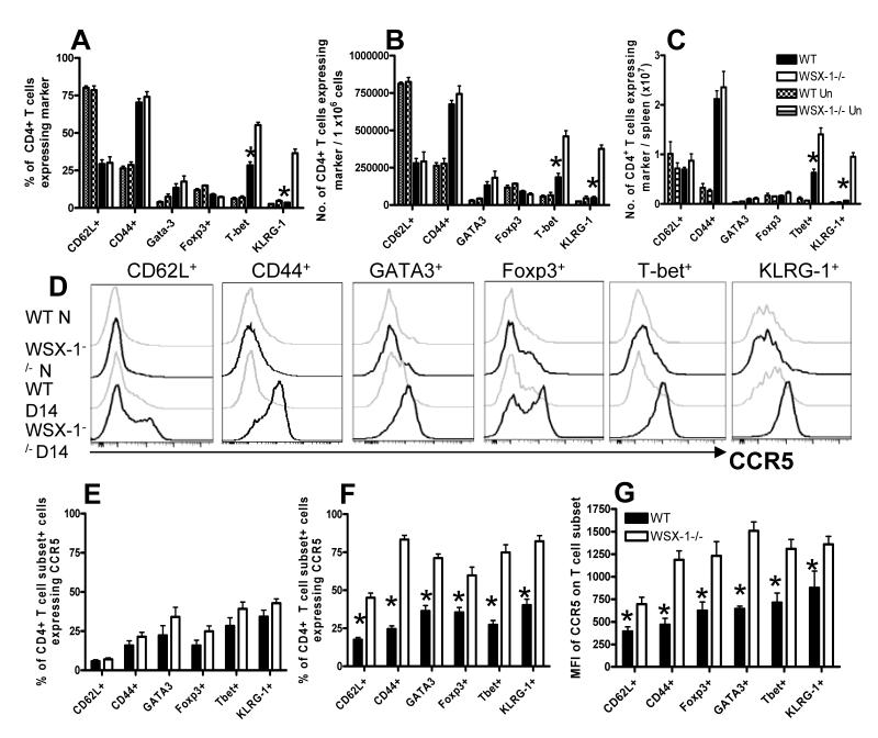
IL-27 exerts pleiotropic suppressive effects on naive and effector T cell populations during infection and inflammation. Surprisingly, however, the role of IL-27 in restricting or shaping effector CD4(+) T cell chemotactic responses, as a mechanism to reduce T cell-dependent tissue inflammation, is unknown. In this study, using Plasmodium berghei NK65 as a model of a systemic, proinflammatory infection, we demonstrate that IL-27R signaling represses chemotaxis of infection-derived splenic CD4(+) T cells in response to the CCR5 ligands, CCL4 and CCL5. Consistent with these observations, CCR5 was expressed on significantly higher frequencies of splenic CD4(+) T cells from malaria-infected, IL-27R-deficient (WSX-1(-/-)) mice than from infected wild-type mice. We find that IL-27 signaling suppresses splenic CD4(+) T cell CCR5-dependent chemotactic responses during infection by restricting CCR5 expression on CD4(+) T cell subtypes, including Th1 cells, and also by controlling the overall composition of the CD4(+) T cell compartment. Diminution of the Th1 response in infected WSX-1(-/-) mice in vivo by neutralization of IL-12p40 attenuated CCR5 expression by infection-derived CD4(+) T cells and also reduced splenic CD4(+) T cell chemotaxis toward CCL4 and CCL5. These data reveal a previously unappreciated role for IL-27 in modulating CD4(+) T cell chemotactic pathways during infection, which is related to its capacity to repress Th1 effector cell development. Thus, IL-27 appears to be a key cytokine that limits the CCR5-CCL4/CCL5 axis during inflammatory settings.


Figures

Figure 1
Figure 1. WSX-1 signalling restricts CD4+ T cell chemotactic responses during malaria infection
WT and WSX-1−/− mice were infected with 104 P. berghei NK65 pRBCs. Splenic CD4+ T cells were purified from naïve and malaria-infected mice (D14 p.i.). (A,B), 1 ×10⁶ purified (A) naïve, or (B) infection-derived, CD4+ T cells were placed in duplicate in the top chamber of a 5μm transwell and migration of cells towards recombinant chemokines (all at 100ng/ml) in the bottom chamber was assessed by microscopy. Data are the mean +/− SEM of the group and are representative of 5 independent experiments. * P<0.05, WT vs WSX-1−/− mice.
Figure 2
Figure 2. SX-1 signalling restricts CCR5 expression on CD4+ T cells during malaria infection
WT and WSX-1−/− mice were infected with 104 P. berghei NK65 pRBCs. (A) Representative plots (gated on CD4+ T cells) showing the surface expression of chemokine receptors on splenic CD4+ T cells from naïve and infected (day 14 p.i.) WT and WSX-1−/− mice. (B, C) frequencies of splenic CD4+ T cells from (B) naïve and (C) malaria infected (day 14 p.i.) WT and WSX-1−/− mice expressing chemokine receptors on the cell surface. (D) The frequencies of splenic CD4+ T cells from malaria infected (day 14 p.i.) WT and WSX-1−/− mice expressing intracellular chemokine receptors. (E) The frequencies of splenic CD4+ T cells from malaria-infected (day 14 p.i.) WT and IL-10R−/− mice expressing chemokine receptors on the cell surface. Data are the mean +/− SEM of the group with 3-5 mice per group. Data are representative of 3 independent experiments. (B-D) * P<0.05, WT vs WSX-1−/− mice. (F) * P<0.05, WT vs IL-10R−/− mice.
Figure 3
Figure 3. Loss of WSX-1 signalling leads to remodelling of the CD4+CCR5+ T cell compartment and increased numbers of Th1-CCR5+ T cells during malaria infection
WT and WSX-1−/− mice were infected with 104 P. berghei NK65 pRBCs. (A) Representative plots showing the surface expression of T cell markers on splenic CD4+ CCR5+ T cells from naïve and infectied (day 14 p.i.) WT and WSX-1−/− mice. (B, C) frequencies and (D, E) numbers of splenic CD4+CCR5+ T cells from (B, D) naïve and (C, E) malaria infected (day 14 p.i.) WT and WSX-1−/− mice expressing the individual markers. (D, E) Numbers of cells calculated out of the 1×10⁶ purified CD4+ T cells used in chemotaxis assays. Data are the mean +/− SEM of the group with 3-5 mice per group and are representative of 4 independent experiments. * P<0.05, WT vs WSX-1−/− mice.
Figure 4
Figure 4. Loss of WSX-1 signalling modifies the structure of the CD4+ T cell population but also leads to unconstrained CCR5 expression on CD4+ T cell subsets during malaria infection
WT and WSX-1−/− mice were infected with 104 P. berghei NK65 pRBCs. (A) Frequencies of splenic CD4+ T cells from naïve and infected (day 14 p.i.) WT and WSX-1−/− mice expressing the various T cell markers. (B, C) The numbers of CD4+ T cells derived from naïve and infection-derived (D14 p.i.) WT and WSX-1−/− mice expressing the various T cell subset markers out of (B) 1 ×10⁶ CD4+ T cells utilised in the chemotaxis assay and (C) total splenocytes. (D) Representative histograms (gated on CD4+ Marker+ T cells) showing the surface expression of CCR5 on splenic CD4+ T cells from naïve and infected (day 14 p.i.) WT and WSX-1−/− mice. (E, F) frequencies and (G) MFI of CCR5 expression on (E) naïve and (F, G) malaria-infection derived (day 14 p.i.) splenic CD4+ Marker+ T cells. Data are the mean +/− SEM of the group with 3-5 mice per group and are representative of 4 independent experiments. * P<0.05, WT vs WSX-1−/− mice.
Figure 5
Figure 5. Coordinated expression of CCR5 with IL-12R on CD4+ T cells derived from malaria-infected WSX-1−/− mice
WT and WSX-1−/− mice were infected with 104 P. berghei NK65 pRBCs. (A) Representative plots (gated on CD4+ T cells) showing the surface expression of CCR5 versus IL-12Rβ1 and CD25 on splenic CD4+ T cells from naïve and infected (day 14 p.i.) WT and WSX-1−/− mice. (B-C) The frequencies of splenic CD4+CCR5+ T cells from naïve and malaria-infected (day 14 p.i.) WT and WSX-1−/− mice expressing (B) IL-12Rβ1 and (C) CD25. Data are the mean +/− SEM of the group with 3-5 mice per group and are representative of 2 independent experiments. * P<0.05, WT vs. WSX-1−/− mice.
Figure 6
Figure 6. Anti-IL-12p40 mAb administration to malaria infected WSX-1−/− mice inhibits T-bet expression, reverses CCR5 expression by CD4+ T cells and attenuates CD4+ T cell chemotaxis
WT and WSX-1−/− mice were infected with 104 P. berghei NK65 pRBCs. Some WSX-1−/− mice were treated with 250μg anti-IL-12p40 or anti-IL-2 mAbs every 2nd day starting on day 7 p.i. (A, B) Representative plots (gated on CD4+ T cells) showing the surface expression of (A) T-bet and (B) CCR5 on splenic CD4+ T cells from naïve and infected (day 14 p.i.) WT and WSX-1−/− mice and WSX-1−/− mice treated with anti-IL-12p40 or anti-IL-2. (C, D) The frequencies of splenic CD4+ T cells from naïve, infected control and infected antibody-treated WT and WSX-1−/− mice expressing (C) T-bet or (D) CCR5. (E) The numbers of CD4+ T cells from WT, anti-IL-12p40 mAb treated and control infected WSX-1−/− mice expressing the various T cell subset markers out of 1 ×10⁶ CD4+ T cells utilised in the chemotaxis assay (F) 1×10⁶ purified CD4+ T cells from infected WT, WSX-1−/− and WSX-1−/− mice treated with anti-IL-12p40 or anti-IL-2 were placed in duplicate in the top chamber of a 5μm transwell and migration towards recombinant CCL4 (100ng/ml) in the bottom chamber was assessed by microscopy. Data are the mean +/− SEM of the group with 3-5 mice per group and are representative of 2 independent experiments. * P<0.05, WT vs WSX-1−/− control mice, ~ P<0.05, WT vs anti-IL-12p40 treated WSX-1−/− mice, + P<0.05, WT vs anti-IL-2 treated WSX-1−/− mice, † P<0.05, WSX-1−/− control mice vs. anti-IL-12p40 treated WSX-1−/− mice, ‡ P<0.05, anti-IL-12p40 treated WSX-1−/− mice vs. anti-IL-2 treated WSX-1−/− mice.

Similar articles

Cited by

References

    1. Yoshida H, Nakaya M, Miyazaki Y. Interleukin 27: a double-edged sword for offense and defense. J Leukoc Biol. 2009;86:1295–303. - PubMed
    1. Hall AO, Silver JS, Hunter CA. The immunobiology of IL-27. Adv Immunol. 2012;115:1–44. - PubMed
    1. Findlay EG, Greig R, Stumhofer JS, Hafalla JC, de Souza JB, Saris CJ, Hunter CA, Riley EM, Couper KN. Essential role for IL-27 receptor signaling in prevention of Th1-mediated immunopathology during malaria infection. J Immunol. 2010;185:2482–92. - PubMed
    1. Villarino A, Hibbert L, Lieberman L, Wilson E, Mak T, Yoshida H, Kastelein RA, Saris C, Hunter CA. The IL-27R (WSX-1) is required to suppress T cell hyperactivity during infection. Immunity. 2003;19:645–55. - PubMed
    1. Hamano S, Himeno K, Miyazaki Y, Ishii K, Yamanaka A, Takeda A, Zhang M, Hisaeda H, Mak TW, Yoshimura A, Yoshida H. WSX-1 is required for resistance to Trypanosoma cruzi infection by regulation of proinflammatory cytokine production. Immunity. 2003;19:657–67. - PubMed

Publication types

MeSH terms