. 2013 May 1;190(9):4861-7.
doi: 10.4049/jimmunol.1202857. Epub 2013 Mar 27.

Circulating fibrocytes prepare the lung for cancer metastasis by recruiting Ly-6C+ monocytes via CCL2

Affiliations

Circulating fibrocytes prepare the lung for cancer metastasis by recruiting Ly-6C+ monocytes via CCL2

Hendrik W van Deventer et al. J Immunol. .

Abstract

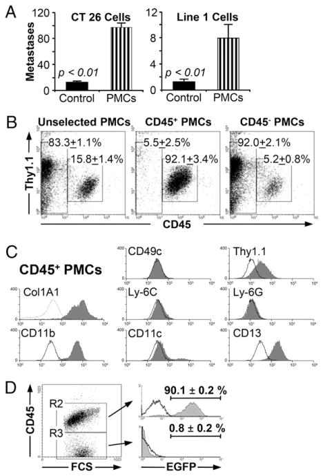
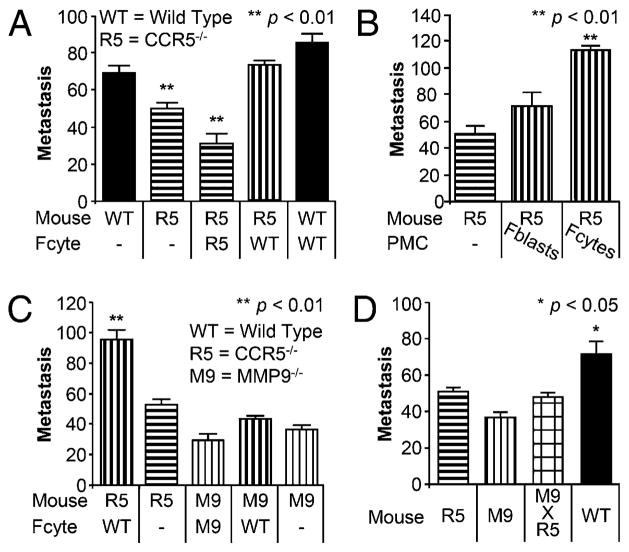
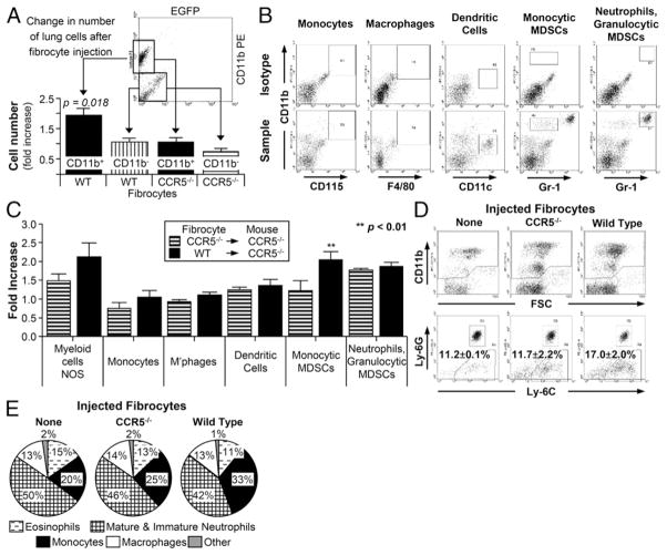
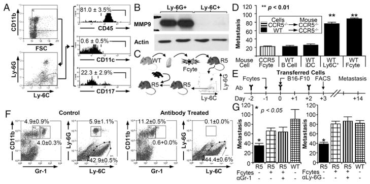
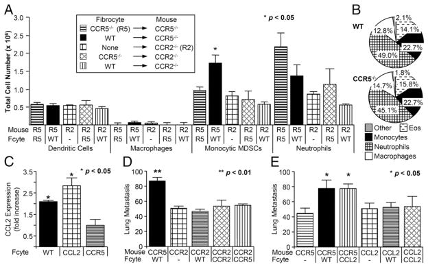
Fibrocytes are circulating, hematopoietic cells that express CD45 and Col1a1. They contribute to wound healing and several fibrosing disorders by mechanisms that are poorly understood. In this report, we demonstrate that fibrocytes predispose the lung to B16-F10 metastasis by recruiting Ly-6C(+) monocytes. To do so, we isolated fibrocytes expressing CD45, CD11b, CD13, and Col1a1 from the lungs of wild type (WT) and Ccr5(-/-) mice. WT but not Ccr5(-/-) fibrocytes increased the number of metastatic foci when injected into Ccr5(-/-) mice (73 ± 2 versus 32 ± 5; p < 0.001). This process was MMP9 dependent. Injection of WT enhanced GFP(+) fibrocytes also increased the number of Gr-1(Int), CD11b(+), and enhanced GFP(-) monocytes. Like premetastatic-niche monocytes, these recruited cells expressed Ly-6C, CD117, and CD45. The transfer of these cells into Ccr5(-/-) mice enhanced metastasis (90 ± 8 foci) compared with B cells (27 ± 2), immature dendritic cells (31 ± 6), or alveolar macrophages (28 ± 3; p < 0.05). WT and Ccl2(-/-) fibrocytes also stimulated Ccl2 expression in the lung by 2.07 ± 0.05- and 2.78 ± 0.36-fold compared with Ccr5(-/-) fibrocytes (1.0 ± 0.06; p < 0.05). Furthermore, WT fibrocytes did not increase Ly-6C(+) monocytes in Ccr2(-/-) mice and did not promote metastasis in either Ccr2(-/-) or Ccl2(-/-) mice. These data support our hypothesis that fibrocytes contribute to premetastatic conditioning by recruiting Ly-6C(+) monocytes in a chemokine-dependent process. This work links metastatic risk to conditions that mobilize fibrocytes, such as inflammation and wound repair.


Conflict of interest statement

Disclosures

The authors have no financial conflicts of interest.

Figures

FIGURE 1
FIGURE 1
Characterization and isolation of fibrocytes. (A) Injection of WT pulmonary mesenchymal cells increases metastases with CT26 and Line 1 tumor cells. The graph shows the number of metastatic foci in BALB/c mice injected with 4 × 10⁵ pulmonary mesenchymal cells (PMCs; striped bars) compared with PBS-injected controls (solid bars). The graph on the left demonstrates a 7.8-fold increase in metastasis after injection with 1 × 10⁵ CT26 cells (n = 20). The graph on the right shows a 6.2-fold increase after an injection with 2 × 10⁵ Line 1 cells (n = 22). (B) CD45+ fibrocytes can be isolated using immunomagnetic bead selection. Pulmonary mesenchymal cells were isolated from minced lung cultures as described. These cells were incubated with CD45 immunomagnetic beads and selected with Miltenyi magnetic columns. Representative dot plots are shown for cells prior to selection (left), CD45 selected cells (center), and flow through cells (right; n = 4). (C) CD45+ PMCs are phenotypically consistent with fibrocytes. Flow cytometry was performed on PMCs for the indicated markers. Representative histograms are shown for CD45+ cells (solid gray) and isotype control (black line). (D) CD45+ fibrocytes originate in the bone marrow. WT bone marrow chimeric mice were formed by injecting 3 × 10⁶ bone marrow cells from EGFP transgenic mice into irradiated WT mice. Six to eight weeks later, the lungs were harvested and PMCs were isolated using the standard 2-wk protocol. The cells were then assessed by flow cytometry. The dot plot on the left shows PMCs characterized by forward scatter and CD45 expression. The histogram on the top right shows the percentage of EGFP+ cells taken from the CD45+ PMCs (R2). The histogram on the bottom shows EGFP+ cells taken from the CD45 PMCs (R3). The unfilled black line is EGFP expression for WT mice injected with WT (non-EGFP) marrow (n = 8).
FIGURE 2
FIGURE 2
WT CD45+ fibrocytes increased metastasis in Ccr5−/− mice via Mmp9. (A) Bar graphs depicting the number of metastases in WT and Ccr5−/− (R5) mice following the injection of 1 × 10⁵ WT or Ccr5−/− fibrocytes (Fcyte); 7.5 × 10⁵ B16-F10 melanoma cells were injected i.v. 24 h after fibrocytes. Metastases were counted 2 wk later (n = 50). (B) Bar graphs depicting metastasis in Ccr5−/− mice (left) and Ccr5−/− mice following injection with either 1 × 10⁵ WT fibroblasts (Fblasts) or fibrocytes (Fcytes; n = 26). (C) Bar graphs showing metastasis in Ccr5−/− and Mmp9−/− (M9) mice following injection with WT or Mmp9−/− fibrocytes (n = 92). (D) Bar graph showing metastasis in mice without fibrocyte injection. WT mice (solid black) had significantly more metastases than Ccr5−/−, Mmp9−/− or the double knockout Mmp9−/− × Ccr5−/− (checkerboard; n = 47).
FIGURE 3
FIGURE 3
WT fibrocytes recruited Ly-6+, Ly-6Glo cells in Ccr5−/− mice. (A) CD11b+ cells were increased in Ccr5−/− mice 24 h after the injection of WT EGFP+ fibrocytes. Dot plot shows gating strategy for calculating CD11b+ cell number. The bar graph shows the fold increases of CD11b+ (black bars) and CD11b (striped; n = 22). (B) Gating strategy for the identification of CD11b+ cells after injection with WT fibrocytes. The top row of dot plots were stained with isotype controls; the bottom row plots were stained with CD115, F4/80, CD11c, and Gr-1. (C) Bar graphs depicting fold induction of cell subpopulations following injection with Ccr5−/− (striped) or WT (solid) fibrocytes. Significant fold induction was seen in only monocytic MDSCs (n = 20). (D) Dot plots from Ccr5−/− mice following the injection of Ccr5−/− (center) or WT (right) fibrocytes. Ly-6C+, Ly-6G were significantly increased after injection of WT cells. (E) Differential percentages of CD11b+ cells as detected on cytospin preparations. All differentials performed on CD11b+ cells from Ccr5−/− mice after injection with WT fibrocytes (left), Ccr5−/− fibrocytes (center) or no fibrocytes (right; n = 24).
FIGURE 4
FIGURE 4
Ly-6C+, Ly-6Glo cells recruited by fibrocytes promoted metastasis. (A) Flow cytometric analysis of Ly-6Glo, Ly-6C+ cells. Gating strategy is on the left; histograms for CD45, CD11c, and CD117 are given on the right (n = 5). (B) Western blot analysis of Ly-6Glo, Ly-6C+ cells isolated from Ccr5−/− mice by flow sorting following injection with WT fibrocytes. (C) Experimental schema for defining function of Ly-6C+ monocytes. Ly-6C+ cells were isolated from Ccr5−/−(R5) mice 24 h after fibrocyte injection and transferred into R5 mice. Tumor cells were injected within 4 h of monocyte injection. (D) Bar graph shows metastasis in Ccr5−/− mice injected with Ccr5−/− fibrocytes or 5 × 10⁶ WT B cells, 1 × 10⁶ WT immature dendritic cells (iDCs), 2 × 10⁵ WT Ly-6C+ monocytes, or WT fibrocytes (n = 42). (E) Experimental schema for the depletion Gr-1 cells. Gr-1 or Ly-6G Ab was given on days −2,, 0, and 2 in relationship to the i.v. injection of B16 F10 cells (day zero). Fibrocytes (1 × 10⁵) were given on day −1; flow cytometry was performed on day 3. (F) Representative dot plots of pulmonary cells from mice treated with either Gr-1 or Ly-6G Ab. The graph on the left includes all cells (excluding debris by FSC/SSC); the graph on the right represents only CD11b+ cells. (G) Bar graph displaying the number of metastasis in mice treated with Gr-1 (n = 15) or Ly-6G (n = 31) Ab. Untreated Ccr5−/− mice (black) had statistically fewer metastases than all other groups. There were no significant differences in Ccr5−/− mice injected with WT fibrocytes.
FIGURE 5
FIGURE 5
Fibrocytes recruited premetastatic monocytes via CCR2. (A) Bar graphs depicting total number of cells for each subpopulation following the injection with Ccr5−/− (R5), Ccr2−/− (R2), or WT fibrocytes. Significant increase was seen in monocytic MDSCs only. (B) Pie graphs showing differential percentages of CD11b+ cells as detected on cytospin preparations. All differentials performed on CD11b+ cells from Ccr2−/− mice after injection with WT (top) or Ccr5−/− fibrocytes (bottom). (C) Bar graph depicting fold increase in Ccl2 mRNA expression in Ccr5−/− mice following injection with WT, Ccl2−/−, or Ccr5−/− fibrocytes (n = 16). (D) Bar graph representing lung metastasis in Ccr5−/− or Ccr2−/− mice injected with WT, Ccr2−/−, or Ccr5−/− fibrocytes (n = 72). (E) Bar graph showing the number of metastases in Ccr5−/− or Ccl2−/− after the injection of WT or Ccl2−/− fibrocytes (Fcyte; n = 70).

Similar articles

Cited by

References

    1. Bucala R, Spiegel LA, Chesney J, Hogan M, Cerami A. Circulating fibrocytes define a new leukocyte subpopulation that mediates tissue repair. Mol Med. 1994;1:71–81. - PMC - PubMed
    1. Bucala R. Review Series – Inflammation & fibrosis fibrocytes and fibrosis. QJM. 2012;105:505–508. - PubMed
    1. Quan TE, Cowper S, Wu SP, Bockenstedt LK, Bucala R. Circulating fibrocytes: collagen-secreting cells of the peripheral blood. Int J Biochem Cell Biol. 2004;36:598–606. - PubMed
    1. Iqbal SA, Sidgwick GP, Bayat A. Identification of fibrocytes from mesenchymal stem cells in keloid tissue: a potential source of abnormal fibroblasts in keloid scarring. Arch Dermatol Res. 2012;304:665–671. - PubMed
    1. Todd JL, Palmer SM. Bronchiolitis obliterans syndrome: the final frontier for lung transplantation. Chest. 2011;140:502–508. - PubMed

Publication types

MeSH terms