. 2013 Jun;87(11):6150-60.
doi: 10.1128/JVI.03372-12. Epub 2013 Mar 27.

TMPRSS2 activates the human coronavirus 229E for cathepsin-independent host cell entry and is expressed in viral target cells in the respiratory epithelium

Affiliations

TMPRSS2 activates the human coronavirus 229E for cathepsin-independent host cell entry and is expressed in viral target cells in the respiratory epithelium

Stephanie Bertram et al. J Virol. 2013 Jun.

Abstract

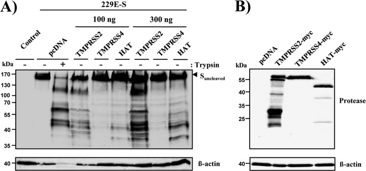
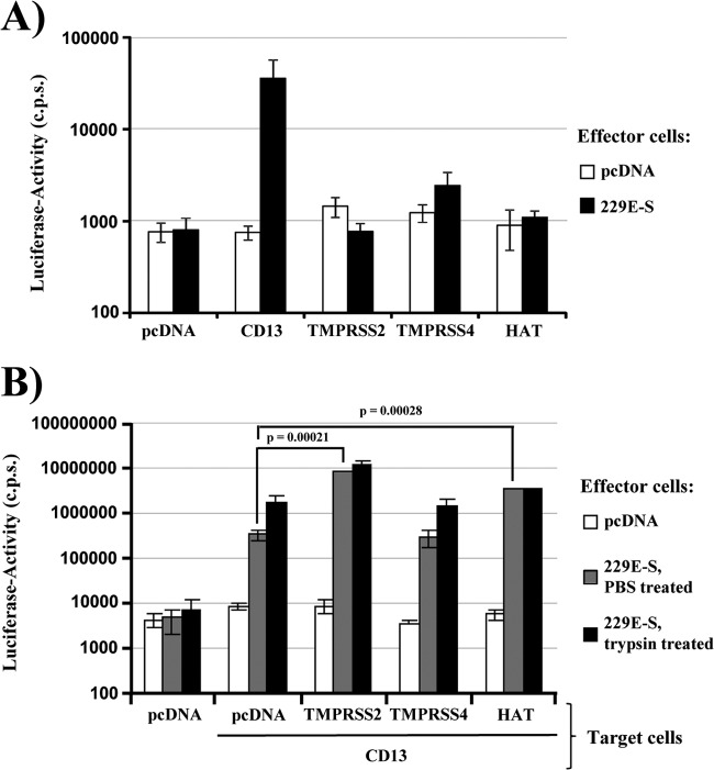
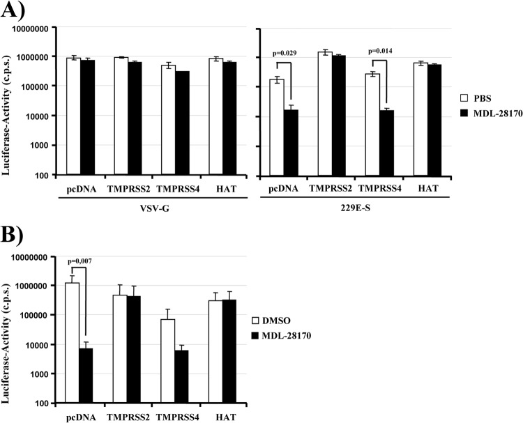
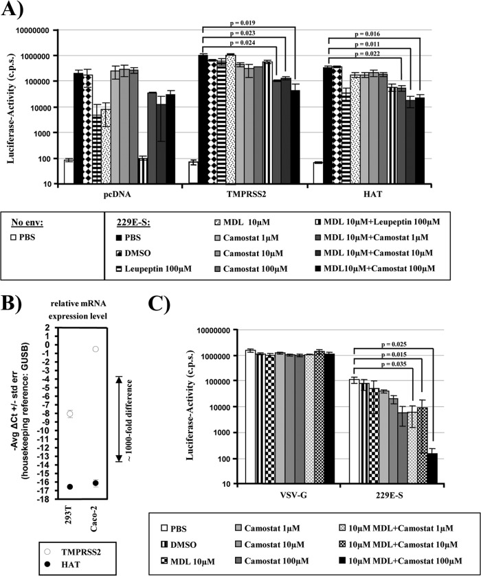
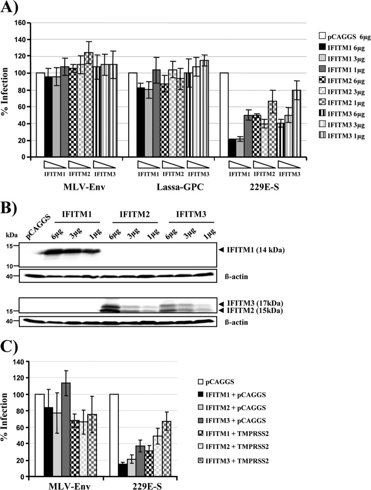
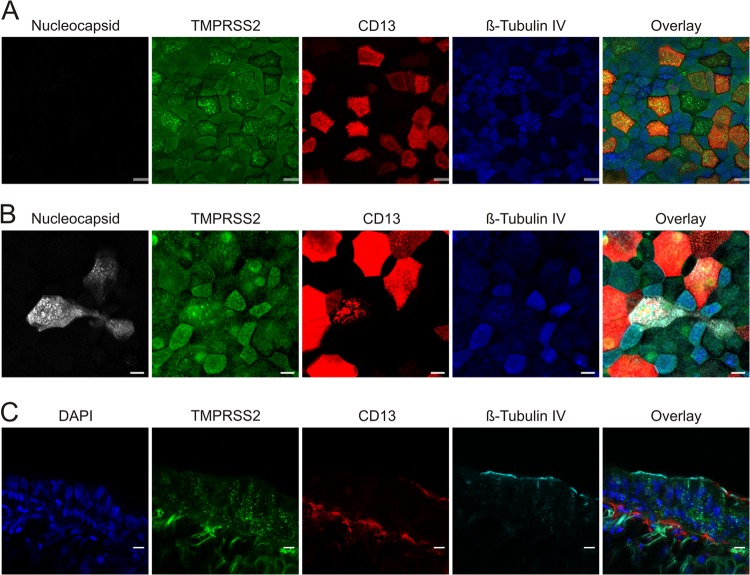
Infection with human coronavirus 229E (HCoV-229E) is associated with the common cold and may result in pneumonia in immunocompromised patients. The viral spike (S) protein is incorporated into the viral envelope and mediates infectious entry of HCoV-229E into host cells, a process that depends on the activation of the S-protein by host cell proteases. However, the proteases responsible for HCoV-229E activation are incompletely defined. Here we show that the type II transmembrane serine proteases TMPRSS2 and HAT cleave the HCoV-229E S-protein (229E-S) and augment 229E-S-driven cell-cell fusion, suggesting that TMPRSS2 and HAT can activate 229E-S. Indeed, engineered expression of TMPRSS2 and HAT rendered 229E-S-driven virus-cell fusion insensitive to an inhibitor of cathepsin L, a protease previously shown to facilitate HCoV-229E infection. Inhibition of endogenous cathepsin L or TMPRSS2 demonstrated that both proteases can activate 229E-S for entry into cells that are naturally susceptible to infection. In addition, evidence was obtained that activation by TMPRSS2 rescues 229E-S-dependent cell entry from inhibition by IFITM proteins. Finally, immunohistochemistry revealed that TMPRSS2 is coexpressed with CD13, the HCoV-229E receptor, in human airway epithelial (HAE) cells, and that CD13(+) TMPRSS2(+) cells are preferentially targeted by HCoV-229E, suggesting that TMPRSS2 can activate HCoV-229E in infected humans. In sum, our results indicate that HCoV-229E can employ redundant proteolytic pathways to ensure its activation in host cells. In addition, our observations and previous work suggest that diverse human respiratory viruses are activated by TMPRSS2, which may constitute a target for antiviral intervention.


Figures

Fig 1
Fig 1
TMPRSS2 and HAT cleave the S-protein of HCoV-229E. (A) An expression plasmid for 229E-S was transiently cotransfected with expression plasmids for the indicated proteases or with empty plasmid (pcDNA) into 293T cells. The cells were harvested and treated with PBS or TPCK-trypsin, as indicated, and 229E-S expression was analyzed by Western blotting employing a goat anti-HCoV-229E serum. The detection of β-actin served as a loading control. (B) Plasmids encoding the indicated TTSPs containing an N-terminal myc tag were transiently transfected into 293T cells. Cells transfected with empty plasmid served as a negative control. Protein expression was detected with anti-myc antibody, and detection of β-actin served as a loading control.
Fig 2
Fig 2
TMPRSS2 and HAT increase cell-cell fusion driven by the S-protein of HCoV-229E. (A) Effector 293T cells were cotransfected with the 229E-S plasmid or empty plasmid (pcDNA), jointly with a plasmid encoding VP16 fused to GAL4. In parallel, 293T target cells were transiently cotransfected with plasmids encoding either human CD13 or the indicated proteases or with empty plasmid, jointly with a plasmid containing a GAL4-VP16-activated luciferase expression cassette. Effector and target cells were mixed, and luciferase activities in cell lysates were determined at 48 h postinfection. The results of a representative experiment performed in triplicate are shown; error bars indicate standard deviations (SD). Similar results were obtained in three separate experiments. (B) Experiments were conducted as described for panel A, but target cells expressing CD13 alone or in conjunction with the indicated proteases were examined. Eight hours after mixing of cells, the effector cell-target cell cocultures were treated with PBS or trypsin for 8 h. The results of a representative experiment performed in triplicate are shown; error bars indicate SD. The results were confirmed in two independent experiments.
Fig 3
Fig 3
TMPRSS2 and HAT allow for cathepsin L-independent host cell entry of HCoV-229E. (A) 293T cells were transiently transfected with plasmids encoding human CD13 and the indicated proteases or with empty plasmid (pcDNA). The cells were seeded into 96-well plates, preincubated with PBS or the cathepsin L inhibitor MDL-28170 (10 μM), and transduced with firefly luciferase-encoding lentiviral vectors bearing VSV-G or 229E-S. Luciferase activities in cell lysates were measured at 72 h postransduction. The results of a representative experiment performed in triplicate are shown; error bars indicate SD. Comparable results were obtained in three independent experiments. (B) Experiments were carried out as described for panel A, but target cells were seeded into 24-well plates, preincubated with 20 μM MDL-28170 or DMSO, and then infected with HCoV-229E encoding Renilla luciferase. Luciferase activities in cell lysates were quantified at 8 h postinfection. The averages for three independent experiments performed in triplicate are shown; error bars indicate SD.
Fig 4
Fig 4
Redundant proteolytic pathways activate the S-protein of HCoV-229E for virus-cell fusion. (A) 293T cells stably expressing CD13 were transiently transfected with plasmids encoding the indicated proteases or with empty plasmid (pcDNA) and were used as target cells for transduction by lentiviral vectors bearing 229E-S. Prior to infection, target cells were incubated with PBS, DMSO, or the indicated protease inhibitors. Luciferase activities in cell lysates were measured at 72 h postinfection. The results of a representative experiment performed in triplicate are shown; error bars indicate SD. Comparable results were observed in three independent experiments. (B) Analysis of TMPRSS2 and HAT expression by quantitative RT-PCR. Total RNAs were prepared from the indicated cell lines, treated with DNase, and reverse transcribed. TMPRSS2, HAT, and GUSB (housekeeping control) were amplified from cDNA by TaqMan gene expression assays. Similar results were obtained in an independent experiment. (C) Caco-2 cells, which express endogenous TMPRSS2, were incubated with PBS, DMSO, or the indicated protease inhibitors, followed by transduction with lentiviral pseudoparticles bearing 229E-S or VSV-G, as a control. At 72 h postinfection, luciferase activities in cell lysates were measured. The results of a representative experiment performed in triplicate are shown; error bars indicate SD. Comparable results were observed in five independent experiments.
Fig 5
Fig 5
TMPRSS2 partially rescues entry driven by the S-protein of HCoV-229E from inhibition by IFITM proteins. (A) 293T-CD13 target cells were transfected with the indicated amounts of IFITM expression plasmids or empty plasmid (pCAGGS) and subsequently transduced with infectivity-normalized pseudotypes carrying the glycoproteins of MLV, Lassa fever virus, or HCoV-229E. Luciferase activities in cell lysates were determined at 72 h postinfection. The results are presented as % infection and represent the averages for three independent experiments; error bars indicate standard errors of the means (SEM). (B) 293T-CD13 cells were transfected with the indicated amounts of expression plasmids encoding IFITM proteins. Cell lysates were prepared after 48 h and analyzed by Western blotting using IFITM-1- and IFITM-2/3-reactive antibodies. (C) 293T-CD13 target cells expressing the indicated IFITM proteins either alone or in combination with TMPRSS2 were transduced with pseudoparticles bearing the glycoprotein of MLV or HCoV-229E, respectively. Cells were processed as described for panel A. The infection efficiency was calculated as % infection based on the infectivity for pCAGGS-transfected target cells, which was set as 100%. Comparable results were obtained in two independent experiments.
Fig 6
Fig 6
TMPRSS2 expression on differentiated HAE cells colocalizes with CD13-expressing cells. The in vitro distribution of TMPRSS2 expression (green) was analyzed in mock (A)- and HCoV-229E (B)-infected HAE cultures that were fixed and immunostained for colocalization with nucleocapsid protein (white) and for CD13 expression (red). To discriminate between ciliated and nonciliated populations, cells were stained with antibodies directed against β-tubulin IV (cilia; blue). Confocal images representative of 4 randomly selected fields from 3 independent experiments are shown. (C) For the in vivo distribution in primary bronchial tissue, TMPRSS2 (green) expression was analyzed in conjunction with CD13 (red), β-tubulin IV/cilia (cyan), and nuclei acids (blue). Confocal images representative of 4 randomly selected fields from 2 independent experiments are shown. Bars, 20 μm.

References

    1. Bermingham A, Chand M, Brown C, Aarons E, Tong C, Langrish C, Hoschler K, Brown K, Galiano M, Myers R, Pebody R, Green H, Boddington N, Gopal R, Price N, Newsholme W, Drosten C, Fouchier R, Zambon M. 2012. Severe respiratory illness caused by a novel coronavirus, in a patient transferred to the United Kingdom from the Middle East, September 2012. Euro Surveill. 17:20290. - PubMed
    1. Corman V, Eckerle I, Bleicker T, Zaki A, Landt O, Eschbach-Bludau M, BSvan Gopal R, Ballhause M, Bestebroer T, Muth D, Muller M, Drexler J, Zambon M, Osterhaus A, Fouchier R, Drosten C. 2012. Detection of a novel human coronavirus by real-time reverse-transcription polymerase chain reaction. Euro Surveill. 17:20285. - PubMed
    1. Drosten C, Gunther S, Preiser W, van der Werf S, Brodt HR, Becker S, Rabenau H, Panning M, Kolesnikova L, Fouchier RA, Berger A, Burguiere AM, Cinatl J, Eickmann M, Escriou N, Grywna K, Kramme S, Manuguerra JC, Muller S, Rickerts V, Sturmer M, Vieth S, Klenk HD, Osterhaus AD, Schmitz H, Doerr HW. 2003. Identification of a novel coronavirus in patients with severe acute respiratory syndrome. N. Engl. J. Med. 348:1967–1976 - PubMed
    1. Ksiazek TG, Erdman D, Goldsmith CS, Zaki SR, Peret T, Emery S, Tong S, Urbani C, Comer JA, Lim W, Rollin PE, Dowell SF, Ling AE, Humphrey CD, Shieh WJ, Guarner J, Paddock CD, Rota P, Fields B, DeRisi J, Yang JY, Cox N, Hughes JM, LeDuc JW, Bellini WJ, Anderson LJ. 2003. A novel coronavirus associated with severe acute respiratory syndrome. N. Engl. J. Med. 348:1953–1966 - PubMed
    1. Osterhaus AD, Fouchier RA, Kuiken T. 2004. The aetiology of SARS: Koch's postulates fulfilled. Philos. Trans. R. Soc. Lond. B Biol. Sci. 359:1081–1082 - PMC - PubMed

Publication types

MeSH terms